 Movie
Movie Controller


+ Open data

- Basic information
| Entry | Database: PDB / ID: 1bb9 | ||||||
|---|---|---|---|---|---|---|---|
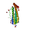
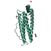
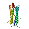
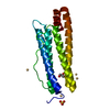
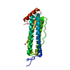
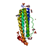
| Title | CRYSTAL STRUCTURE OF THE SH3 DOMAIN FROM RAT AMPHIPHYSIN 2 | ||||||
 Components Components | AMPHIPHYSIN 2 | ||||||
 Keywords Keywords | TRANSFERASE / SH3 DOMAIN | ||||||
| Function / homology |  Function and homology information Function and homology informationregulation of cell cycle => GO:0051726 / lipid tube / negative regulation of ventricular cardiac muscle cell action potential / negative regulation of calcium ion transmembrane transport via high voltage-gated calcium channel / negative regulation of aspartic-type endopeptidase activity involved in amyloid precursor protein catabolic process / lipid tube assembly / T-tubule organization / muscle cell differentiation / negative regulation of potassium ion transmembrane transport / varicosity ...regulation of cell cycle => GO:0051726 / lipid tube / negative regulation of ventricular cardiac muscle cell action potential / negative regulation of calcium ion transmembrane transport via high voltage-gated calcium channel / negative regulation of aspartic-type endopeptidase activity involved in amyloid precursor protein catabolic process / lipid tube assembly / T-tubule organization / muscle cell differentiation / negative regulation of potassium ion transmembrane transport / varicosity / extrinsic component of synaptic vesicle membrane / axon initial segment / cerebellar mossy fiber / positive regulation of astrocyte differentiation / Clathrin-mediated endocytosis / nucleus localization / node of Ranvier / aspartic-type endopeptidase inhibitor activity / I band / RNA polymerase binding / nucleus organization / regulation of neuron differentiation / endosome to lysosome transport / negative regulation of amyloid-beta formation / positive regulation of endocytosis / positive regulation of actin filament polymerization / regulation of heart rate by cardiac conduction / synaptic vesicle endocytosis / axon terminus / T-tubule / phospholipid binding / tau protein binding / Z disc / positive regulation of GTPase activity / actin filament binding / synaptic vesicle / GTPase binding / nuclear envelope / protein-folding chaperone binding / protease binding / microtubule / endosome / positive regulation of apoptotic process / axon / glutamatergic synapse / synapse / protein-containing complex binding / membrane / identical protein binding / nucleus / plasma membraneSimilarity search - Function | ||||||
| Biological species |  Rattus norvegicus (Norway rat) Rattus norvegicus (Norway rat) | ||||||
| Method | X-RAY DIFFRACTION / SYNCHROTRON / SIR / Resolution: 2.2 Å | ||||||
 Authors Authors | Owen, D.J. / Mcmahon, H.T. / Evans, P.R. | ||||||
 Citation Citation | Journal: EMBO J. / Year: 1998 Title: Crystal structure of the amphiphysin-2 SH3 domain and its role in the prevention of dynamin ring formation. Authors: Owen, D.J. / Wigge, P. / Vallis, Y. / Moore, J.D. / Evans, P.R. / McMahon, H.T. | ||||||
| History |
|
- Structure visualization
| Structure viewer | Molecule: MolmilJmol/JSmol |
|---|
- Downloads & links
-Download
| PDBx/mmCIF format | 1bb9.cif.gz | 31.1 KB | Display | PDBx/mmCIF format |
|---|---|---|---|---|
| PDB format | pdb1bb9.ent.gz | 20.1 KB | Display | PDB format |
| PDBx/mmJSON format | 1bb9.json.gz | Tree view | PDBx/mmJSON format | |
| Others |  Other downloads Other downloads |
-Validation report
| Arichive directory | https://data.pdbj.org/pub/pdb/validation_reports/bb/1bb9ftp://data.pdbj.org/pub/pdb/validation_reports/bb/1bb9 | HTTPS FTP |
|---|
-Related structure data
| Similar structure data |
|---|


-Links
 PDBj
PDBj

- Assembly
| Deposited unit | 
| ||||||||
|---|---|---|---|---|---|---|---|---|---|
| 1 | 
| ||||||||
| Unit cell |
|
-Components
| #1: Protein | Mass: 12879.215 Da / Num. of mol.: 1 / Fragment: SH3 DOMAIN Source method: isolated from a genetically manipulated source Source: (gene. exp.) Rattus norvegicus (Norway rat) / Organ: BRAIN / Plasmid: PET15B / Species (production host): Escherichia coli / Production host:  Escherichia coli BL21 (bacteria) / Strain (production host): BL21 / References: UniProt: O08839 Escherichia coli BL21 (bacteria) / Strain (production host): BL21 / References: UniProt: O08839 |
|---|---|
| #2: Water | ChemComp-HOH / Water Mass: 18.015 Da / Num. of mol.: 60 / Source method: isolated from a natural source / Formula: H2O Mass: 18.015 Da / Num. of mol.: 60 / Source method: isolated from a natural source / Formula: H2O |
| Compound details | THIS IS THE SH3 DOMAIN AT THE C-TERMINUS OF AMPHIPHYSIN-2. THIS DOMAIN BINDS TO THE PROLINE-RICH C- ...THIS IS THE SH3 DOMAIN AT THE C-TERMINUS OF AMPHIPHYSI |
-Experimental details
-Experiment
| Experiment | Method: X-RAY DIFFRACTION / Number of used crystals: 2 |
|---|
- Sample preparation
| Crystal | Density Matthews: 2.01 Å3/Da / Density % sol: 38.95 % Description: NATIVE DATA WERE MERGED FROM 2 CRYSTALS, ONE SET TO 2.2 ANGSTROM RESOLUTION COLLECTED ON THE SRS SYNCHROTRON PX9.5, THE OTHER COLLECTED TO 3.5 ANGSTROMS WAS COLLECTED ON A ROTATING ANODE. | |||||||||||||||||||||||||
|---|---|---|---|---|---|---|---|---|---|---|---|---|---|---|---|---|---|---|---|---|---|---|---|---|---|---|
| Crystal grow | Method: vapor diffusion / pH: 6 Details: VAPOUR DIFFUSION, PROTEIN 30MG/ML, RESERVOIR 2.0M AMMONIUM SULFATE, 100MM NA CITRATE PH6.0, PLUS 2MM DTT., vapor diffusion | |||||||||||||||||||||||||
| Crystal grow | *PLUS Temperature: 16 ℃ / Method: vapor diffusion, hanging drop | |||||||||||||||||||||||||
| Components of the solutions | *PLUS
|
-Data collection
| Diffraction | Mean temperature: 293 K |
|---|---|
| Diffraction source | Source: SYNCHROTRON / Site: SRS  / Beamline: PX9.5 / Wavelength: 0.9 / Beamline: PX9.5 / Wavelength: 0.9 |
| Detector | Type: MARRESEARCH / Detector: IMAGE PLATE / Date: Apr 1, 1997 |
| Radiation | Monochromatic (M) / Laue (L): M / Scattering type: x-ray |
| Radiation wavelength | Wavelength: 0.9 Å / Relative weight: 1 |
| Reflection | Resolution: 2.2→36 Å / Num. obs: 5401 / % possible obs: 99 % / Observed criterion σ(I): 6 / Redundancy: 4.2 % / Biso Wilson estimate: 35 Å2 / Rmerge(I) obs: 0.093 / Rsym value: 0.093 / Net I/σ(I): 4.7 |
| Reflection shell | Resolution: 2.2→2.32 Å / Redundancy: 3.2 % / Rmerge(I) obs: 0.201 / Mean I/σ(I) obs: 3.4 / Rsym value: 0.201 / % possible all: 99 |
| Reflection | *PLUS % possible obs: 99.2 % |
| Reflection shell | *PLUS % possible obs: 99.7 % |
- Processing
| Software |
| ||||||||||||||||||||||||||||||||||||||||||||||||||||||||||||||||||||||||||||||||||||
|---|---|---|---|---|---|---|---|---|---|---|---|---|---|---|---|---|---|---|---|---|---|---|---|---|---|---|---|---|---|---|---|---|---|---|---|---|---|---|---|---|---|---|---|---|---|---|---|---|---|---|---|---|---|---|---|---|---|---|---|---|---|---|---|---|---|---|---|---|---|---|---|---|---|---|---|---|---|---|---|---|---|---|---|---|---|
| Refinement | Method to determine structure: SIR / Resolution: 2.2→36 Å / Cross valid method: THROUGHOUT / σ(F): 0
| ||||||||||||||||||||||||||||||||||||||||||||||||||||||||||||||||||||||||||||||||||||
| Displacement parameters | Biso mean: 39 Å2 | ||||||||||||||||||||||||||||||||||||||||||||||||||||||||||||||||||||||||||||||||||||
| Refine analyze | Luzzati sigma a obs: 0.24 Å | ||||||||||||||||||||||||||||||||||||||||||||||||||||||||||||||||||||||||||||||||||||
| Refinement step | Cycle: LAST / Resolution: 2.2→36 Å
| ||||||||||||||||||||||||||||||||||||||||||||||||||||||||||||||||||||||||||||||||||||
| Refine LS restraints |
| ||||||||||||||||||||||||||||||||||||||||||||||||||||||||||||||||||||||||||||||||||||
| Software | *PLUS Name: REFMAC / Classification: refinement | ||||||||||||||||||||||||||||||||||||||||||||||||||||||||||||||||||||||||||||||||||||
| Refinement | *PLUS Rfactor obs: 0.19 / Rfactor Rfree: 0.258 | ||||||||||||||||||||||||||||||||||||||||||||||||||||||||||||||||||||||||||||||||||||
| Solvent computation | *PLUS | ||||||||||||||||||||||||||||||||||||||||||||||||||||||||||||||||||||||||||||||||||||
| Displacement parameters | *PLUS |
