 Movie
Movie Controller

[English] 日本語
 Yorodumi
- PDB-1b7v: Structure of the C-553 cytochrome from Bacillus pasteruii to 1.7 ... -
+ Open data

- Basic information
| Entry | Database: PDB / ID: 1b7v | |||||||||
|---|---|---|---|---|---|---|---|---|---|---|
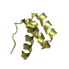
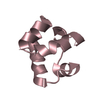
| Title | Structure of the C-553 cytochrome from Bacillus pasteruii to 1.7 A resolution | |||||||||
 Components Components | PROTEIN (CYTOCHROME C-553) | |||||||||
 Keywords Keywords |  ELECTRON TRANSFER / CYTOCHROME ELECTRON TRANSFER / CYTOCHROME | |||||||||
| Function / homology |  Function and homology informationelectron transfer activity / iron ion binding / heme binding / plasma membrane Function and homology informationelectron transfer activity / iron ion binding / heme binding / plasma membraneSimilarity search - Function | |||||||||
| Biological species |  Sporosarcina pasteurii (bacteria) Sporosarcina pasteurii (bacteria) | |||||||||
| Method | X-RAY DIFFRACTION / SYNCHROTRON / MAD / Resolution: 1.7 Å | |||||||||
 Authors Authors | Gonzalez, A. / Benini, S. / Rypniewski, W.R. / Wilson, K.S. / Ciurli, S. | |||||||||
 Citation Citation | Journal: Biochemistry / Year: 2000 Title: Crystal structure of oxidized Bacillus pasteurii cytochrome c553 at 0.97-A resolution. Authors: Benini, S. / Gonzalez, A. / Rypniewski, W.R. / Wilson, K.S. / Van Beeumen, J.J. / Ciurli, S. #1: Journal: J.Biol.Inorg.Chem. / Year: 1998Title: Modulation of Bacillus Pasteurii Cytochrome C553 Reduction Potential by Structural and Solution Parameters Authors: Benini, S. / Borsari, M. / Ciurli, S. / Dikiy, A. / Lamborghini, M. #2: Journal: Proteins / Year: 1997Title: Crystals of Cytochrome C-553 from Bacillus Pasteurii Show Diffraction to 0.97 A Resolution Authors: Benini, S. / Ciurli, S. / Rypniewski, W.R. / Wilson, K. | |||||||||
| History |
|
- Structure visualization
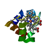
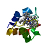
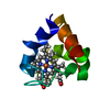
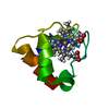
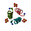
| Structure viewer | Molecule: MolmilJmol/JSmol |
|---|
- Downloads & links
-Download
| PDBx/mmCIF format | 1b7v.cif.gz | 30.4 KB | Display | PDBx/mmCIF format |
|---|---|---|---|---|
| PDB format | pdb1b7v.ent.gz | 19.1 KB | Display | PDB format |
| PDBx/mmJSON format | 1b7v.json.gz | Tree view | PDBx/mmJSON format | |
| Others |  Other downloads Other downloads |
-Validation report
| Arichive directory | https://data.pdbj.org/pub/pdb/validation_reports/b7/1b7vftp://data.pdbj.org/pub/pdb/validation_reports/b7/1b7v | HTTPS FTP |
|---|
-Related structure data


-Links
 PDBj
PDBj


- Assembly
| Deposited unit | 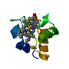
| ||||||||||
|---|---|---|---|---|---|---|---|---|---|---|---|
| 1 |
| ||||||||||
| Unit cell |
|
-Components
| #1: Protein | Mass: 7115.937 Da / Num. of mol.: 1 / Source method: isolated from a natural source / Details: GERMAN COLLECTION OF MICROORGANISMS (DSM) / Source: (natural) Sporosarcina pasteurii (bacteria) / Cellular location: MEMBRANE-BOUNDBiological membrane / Strain: 33 / References: UniProt: P82599 |
|---|---|
| #2: Chemical | ChemComp-HEC / Heme C  Mass: 618.503 Da / Num. of mol.: 1 / Source method: obtained synthetically / Formula: C34H34FeN4O4 Mass: 618.503 Da / Num. of mol.: 1 / Source method: obtained synthetically / Formula: C34H34FeN4O4 |
| #3: Water | ChemComp-HOH / Water Mass: 18.015 Da / Num. of mol.: 128 / Source method: isolated from a natural source / Formula: H2O Mass: 18.015 Da / Num. of mol.: 128 / Source method: isolated from a natural source / Formula: H2O |
| Sequence details | TER LYS: THE FIRST 21 RESIDUES WERE NOT VISIBLE, PROTEIN PRESUMEDLY |
-Experimental details
-Experiment
| Experiment | Method: X-RAY DIFFRACTION / Number of used crystals: 1 |
|---|
- Sample preparation
| Crystal | Density Matthews: 2.1 Å3/Da / Density % sol: 41 % | |||||||||||||||||||||||||
|---|---|---|---|---|---|---|---|---|---|---|---|---|---|---|---|---|---|---|---|---|---|---|---|---|---|---|
| Crystal grow | Temperature: 293 K / Method: vapor diffusion, hanging drop / pH: 8 Details: 8MG/ML OF CYTOCHROME, 20MM TRIS.HCL, PH 8.0 AT 20 DEGREES C, HANGING DROPS IN HAMPTON RESEARCH 24-WELL LINBRO PLATES, VAPOR DIFFUSION, HANGING DROP, temperature 293K | |||||||||||||||||||||||||
| Components of the solutions | Name: TRIS.HCL | |||||||||||||||||||||||||
| Crystal | *PLUS Density % sol: 41 % | |||||||||||||||||||||||||
| Crystal grow | *PLUS Temperature: 20 K | |||||||||||||||||||||||||
| Components of the solutions | *PLUS
|
-Data collection
| Diffraction |
| ||||||||||||||||||||
|---|---|---|---|---|---|---|---|---|---|---|---|---|---|---|---|---|---|---|---|---|---|
| Diffraction source |
| ||||||||||||||||||||
| Detector | Type: MARRESEARCH / Detector: IMAGE PLATE / Date: Dec 15, 1996 / Details: MIRRORS | ||||||||||||||||||||
| Radiation | Monochromator: SI(111) / Protocol: MAD / Monochromatic (M) / Laue (L): M / Scattering type: x-ray | ||||||||||||||||||||
| Radiation wavelength |
| ||||||||||||||||||||
| Reflection | Resolution: 1.7→20 Å / Num. obs: 7404 / % possible obs: 99.5 % / Redundancy: 3.5 % / Biso Wilson estimate: 10.3 Å2 / Rsym value: 0.04 / Net I/σ(I): 8.6 | ||||||||||||||||||||
| Reflection shell | Resolution: 1.7→1.76 Å / Redundancy: 3.5 % / Mean I/σ(I) obs: 5.4 / Rsym value: 0.101 / % possible all: 97.1 | ||||||||||||||||||||
| Reflection | *PLUS Redundancy: 3.52 % / Num. measured all: 26088 / Rmerge(I) obs: 0.04 | ||||||||||||||||||||
| Reflection shell | *PLUS Highest resolution: 1.7 Å / % possible obs: 97.1 % |

- Processing
| Software |
| ||||||||||||||||||||||||||||||||||||||||||||||||||||||||||||||||||||||||||||||||||||
|---|---|---|---|---|---|---|---|---|---|---|---|---|---|---|---|---|---|---|---|---|---|---|---|---|---|---|---|---|---|---|---|---|---|---|---|---|---|---|---|---|---|---|---|---|---|---|---|---|---|---|---|---|---|---|---|---|---|---|---|---|---|---|---|---|---|---|---|---|---|---|---|---|---|---|---|---|---|---|---|---|---|---|---|---|---|
| Refinement | Method to determine structure: MAD / Resolution: 1.7→20 Å / SU B: 1.94 / SU ML: 0.06 / σ(F): 0 / ESU R: 0.12 / ESU R Free: 0.11
| ||||||||||||||||||||||||||||||||||||||||||||||||||||||||||||||||||||||||||||||||||||
| Displacement parameters | Biso mean: 10.3 Å2 | ||||||||||||||||||||||||||||||||||||||||||||||||||||||||||||||||||||||||||||||||||||
| Refinement step | Cycle: LAST / Resolution: 1.7→20 Å
| ||||||||||||||||||||||||||||||||||||||||||||||||||||||||||||||||||||||||||||||||||||
| Refine LS restraints |
| ||||||||||||||||||||||||||||||||||||||||||||||||||||||||||||||||||||||||||||||||||||
| Software | *PLUS Name: REFMAC / Classification: refinement | ||||||||||||||||||||||||||||||||||||||||||||||||||||||||||||||||||||||||||||||||||||
| Refinement | *PLUS Highest resolution: 1.7 Å / σ(F): 0 / % reflection Rfree: 5 % | ||||||||||||||||||||||||||||||||||||||||||||||||||||||||||||||||||||||||||||||||||||
| Solvent computation | *PLUS | ||||||||||||||||||||||||||||||||||||||||||||||||||||||||||||||||||||||||||||||||||||
| Displacement parameters | *PLUS Biso mean: 10.3 Å2 | ||||||||||||||||||||||||||||||||||||||||||||||||||||||||||||||||||||||||||||||||||||
| Refine LS restraints | *PLUS
|
