 Movie
Movie Controller


+ Open data

- Basic information
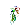
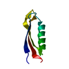
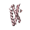
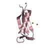
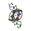
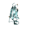
| Entry | Database: PDB / ID: 1b4r | ||||||
|---|---|---|---|---|---|---|---|
| Title | PKD DOMAIN 1 FROM HUMAN POLYCYSTEIN-1 | ||||||
 Components Components | PROTEIN (PKD1_HUMAN) | ||||||
 Keywords Keywords |  MEMBRANE PROTEIN / PKD DOMAIN 1 FROM HUMAN POLYCYSTEIN-1 / POLYCYSTIN (PRECURSOR) MEMBRANE PROTEIN / PKD DOMAIN 1 FROM HUMAN POLYCYSTEIN-1 / POLYCYSTIN (PRECURSOR) | ||||||
| Function / homology |  Function and homology information Function and homology informationmetanephric distal tubule morphogenesis / polycystin complex / mesonephric tubule development / mesonephric duct development / : / metanephric ascending thin limb development / lung epithelium development / metanephric proximal tubule development / lymph vessel morphogenesis / mitocytosis ...metanephric distal tubule morphogenesis / polycystin complex / mesonephric tubule development / mesonephric duct development / : / metanephric ascending thin limb development / lung epithelium development / metanephric proximal tubule development / lymph vessel morphogenesis / mitocytosis / migrasome / calcium-independent cell-matrix adhesion / Wnt receptor activity / VxPx cargo-targeting to cilium / establishment of epithelial cell polarity / genitalia development / detection of mechanical stimulus / response to fluid shear stress / cation channel complex / placenta blood vessel development / Golgi-associated vesicle membrane / digestive tract development / metanephric collecting duct development / transcription regulator inhibitor activity / motile cilium / cartilage development / neural tube development / protein heterotetramerization / ciliary membrane / skin development / cartilage condensation / spinal cord development / branching morphogenesis of an epithelial tube / homophilic cell adhesion via plasma membrane adhesion molecules / regulation of G1/S transition of mitotic cell cycle / cell surface receptor signaling pathway via JAK-STAT / lateral plasma membrane / anatomical structure morphogenesis / embryonic placenta development / regulation of proteasomal protein catabolic process / regulation of cell adhesion / regulation of mitotic spindle organization / calcium channel complex / protein export from nucleus / cell-matrix adhesion / liver development / kidney development / calcium ion transmembrane transport / calcium channel activity / cilium / calcium ion transport / heart development / positive regulation of cytosolic calcium ion concentration / carbohydrate binding / basolateral plasma membrane / peptidyl-serine phosphorylation / in utero embryonic development / transmembrane transporter binding / regulation of cell cycle / protein domain specific binding / Golgi membrane / protein kinase binding / Golgi apparatus / cell surface / endoplasmic reticulum / positive regulation of transcription by RNA polymerase II / extracellular exosome / membrane / nucleus / plasma membrane / cytoplasmSimilarity search - Function | ||||||
| Biological species |  Homo sapiens (human) Homo sapiens (human) | ||||||
| Method | SOLUTION NMR / SA | ||||||
 Authors Authors | Bycroft, M. | ||||||
 Citation Citation | Journal: EMBO J. / Year: 1999 Title: The structure of a PKD domain from polycystin-1: implications for polycystic kidney disease. Authors: Bycroft, M. / Bateman, A. / Clarke, J. / Hamill, S.J. / Sandford, R. / Thomas, R.L. / Chothia, C. | ||||||
| History |
|
- Structure visualization
| Structure viewer | Molecule: MolmilJmol/JSmol |
|---|
- Downloads & links
-Download
| PDBx/mmCIF format | 1b4r.cif.gz | 430.1 KB | Display | PDBx/mmCIF format |
|---|---|---|---|---|
| PDB format | pdb1b4r.ent.gz | 373.4 KB | Display | PDB format |
| PDBx/mmJSON format | 1b4r.json.gz | Tree view | PDBx/mmJSON format | |
| Others |  Other downloads Other downloads |
-Validation report
| Arichive directory | https://data.pdbj.org/pub/pdb/validation_reports/b4/1b4rftp://data.pdbj.org/pub/pdb/validation_reports/b4/1b4r | HTTPS FTP |
|---|
-Related structure data
| Similar structure data |
|---|


-Links
 PDBj
PDBj


- Assembly
| Deposited unit | 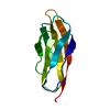
| |||||||||
|---|---|---|---|---|---|---|---|---|---|---|
| 1 |
| |||||||||
| NMR ensembles |
|
-Components
| #1: Protein | Mass: 7963.906 Da / Num. of mol.: 1 / Fragment: PKD DOMAIN Source method: isolated from a genetically manipulated source Source: (gene. exp.) Homo sapiens (human) / Plasmid: PRSETA / Species (production host): Escherichia coli / Production host:  Escherichia coli BL21(DE3) (bacteria) / Strain (production host): BL21(DE3) / References: UniProt: P98161 Escherichia coli BL21(DE3) (bacteria) / Strain (production host): BL21(DE3) / References: UniProt: P98161 |
|---|
-Experimental details
-Experiment
| Experiment | Method: SOLUTION NMR |
|---|
- Sample preparation
| Sample conditions | pH: 3.0 / Temperature: 298 K |
|---|---|
| Crystal grow | *PLUS Method: other / Details: NMR |
-NMR measurement
| NMR spectrometer | Type: Bruker AMX 500 / Manufacturer: Bruker / Model: AMX 500 / Field strength: 500 MHz |
|---|
- Processing
| NMR software |
| ||||||||||||
|---|---|---|---|---|---|---|---|---|---|---|---|---|---|
| Refinement | Method: SA / Software ordinal: 1 | ||||||||||||
| NMR ensemble | Conformer selection criteria: LEAST RESTRAINT VIOLATION / Conformers calculated total number: 50 / Conformers submitted total number: 20 |
