 Movie
Movie Controller

[English] 日本語
 Yorodumi
- PDB-1b3i: NMR SOLUTION STRUCTURE OF PLASTOCYANIN FROM THE PHOTOSYNTHETIC PR... -
+ Open data

- Basic information
| Entry | Database: PDB / ID: 1b3i | ||||||
|---|---|---|---|---|---|---|---|
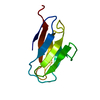
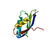
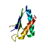
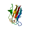
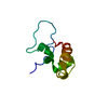
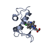
| Title | NMR SOLUTION STRUCTURE OF PLASTOCYANIN FROM THE PHOTOSYNTHETIC PROKARYOTE, PROCHLOROTHRIX HOLLANDICA (MINIMIZED AVERAGE STRUCTURE) | ||||||
 Components Components | PROTEIN (PLASTOCYANIN) | ||||||
 Keywords Keywords |  ELECTRON TRANSPORT / TYPE I COPPER PROTEIN / PHOTOSYNTHESIS ELECTRON TRANSPORT / TYPE I COPPER PROTEIN / PHOTOSYNTHESIS | ||||||
| Function / homology |  Function and homology information Function and homology informationplasma membrane-derived thylakoid membrane / electron transfer activity / copper ion bindingSimilarity search - Function | ||||||
| Biological species |  Prochlorothrix hollandica (bacteria) Prochlorothrix hollandica (bacteria) | ||||||
| Method | SOLUTION NMR / torsion angle dynamics | ||||||
 Authors Authors | Babu, C.R. / Volkman, B.F. / Bullerjahn, G.S. | ||||||
 Citation Citation | Journal: Biochemistry / Year: 1999 Title: NMR solution structure of plastocyanin from the photosynthetic prokaryote, Prochlorothrix hollandica. Authors: Babu, C.R. / Volkman, B.F. / Bullerjahn, G.S. | ||||||
| History |
|
- Structure visualization
| Structure viewer | Molecule: MolmilJmol/JSmol |
|---|
- Downloads & links
-Download
| PDBx/mmCIF format | 1b3i.cif.gz | 40.2 KB | Display | PDBx/mmCIF format |
|---|---|---|---|---|
| PDB format | pdb1b3i.ent.gz | 27.1 KB | Display | PDB format |
| PDBx/mmJSON format | 1b3i.json.gz | Tree view | PDBx/mmJSON format | |
| Others |  Other downloads Other downloads |
-Validation report
| Arichive directory | https://data.pdbj.org/pub/pdb/validation_reports/b3/1b3iftp://data.pdbj.org/pub/pdb/validation_reports/b3/1b3i | HTTPS FTP |
|---|
-Related structure data


-Links
 PDBj
PDBj- Assembly
| Deposited unit | 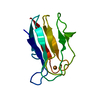
| |||||||||
|---|---|---|---|---|---|---|---|---|---|---|
| 1 |
| |||||||||
| NMR ensembles |
|
-Components
| #1: Protein | Mass: 10148.560 Da / Num. of mol.: 1 / Mutation: T2S Source method: isolated from a genetically manipulated source Details: T2S MUTATION INTRODUCED TO CLONE IN EXPRESSION VECTOR Source: (gene. exp.) Prochlorothrix hollandica (bacteria) / Description: INCLUSION BODIES WERE REFOLDED IN-VITRO / Cellular location: THYLAKOID LUMENThylakoid / Gene: PETE / Plasmid: PVAPC10Cellular location (production host): CYTOPLASM - INCLUSION BODIES Gene (production host): PETE / Production host: Escherichia coli (E. coli) / Strain (production host): BL21(DE3)PLYSS / References: UniProt: P50057, EC: 1.10.99.1 |
|---|---|
| #2: Chemical | ChemComp-CU1 / Copper  Mass: 63.546 Da / Num. of mol.: 1 / Source method: obtained synthetically / Formula: Cu Mass: 63.546 Da / Num. of mol.: 1 / Source method: obtained synthetically / Formula: Cu |
| Sequence details | T2S - INTRODUCED |
-Experimental details
-Experiment
| Experiment | Method: SOLUTION NMR | ||||||||||||||||
|---|---|---|---|---|---|---|---|---|---|---|---|---|---|---|---|---|---|
| NMR experiment |
| ||||||||||||||||
| NMR details | Text: MINIMIZED AVERAGE STRUCTURE. THE STRUCTURE WAS DETERMINED USING TWO- DIMENCIONAL NMR SPECTROSCOPY. WATER SUPPRESSION WAS ACHIEVED WITH A WATERGATE SEQ. WITH A 3-9-19 SELECTIVE INVERSION. |
- Sample preparation
| Details | Contents: 90% WATER/10% D2O |
|---|---|
| Sample conditions | Ionic strength: 20mM POTASSIUM PHOSPHATE / pH: 7.0 / Pressure: 1 atm / Temperature: 298 K |
| Crystal grow | *PLUS Method: other / Details: NMR |
-NMR measurement
| NMR spectrometer | Type: Bruker DMX750 / Manufacturer: Bruker / Model: DMX750 / Field strength: 750 MHz |
|---|
- Processing
| NMR software |
| ||||||||||||||||
|---|---|---|---|---|---|---|---|---|---|---|---|---|---|---|---|---|---|
| Refinement | Method: torsion angle dynamics / Software ordinal: 1 Details: REFINEMENT DETAILS ARE IN THE SUBMITTED FILE AND IN THE JRNL CITATION ABOVE. THE RESTRAINTS ARE IN THE FILES WITH THE 19 CONFORMERS. | ||||||||||||||||
| NMR ensemble | Conformer selection criteria: LOWEST TARGET FUNCTION / Conformers calculated total number: 40 / Conformers submitted total number: 1 |
