 Movie
Movie Controller


+ Open data

- Basic information
| Entry | Database: PDB / ID: 1avc | ||||||
|---|---|---|---|---|---|---|---|
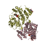
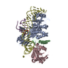
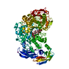
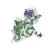
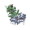
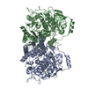
| Title | BOVINE ANNEXIN VI (CALCIUM-BOUND) | ||||||
 Components Components | ANNEXIN VI | ||||||
 Keywords Keywords | CALCIUM/PHOSPHOLIPID-BINDING PROTEIN / ANNEXIN / CALCIUM-BINDING / MEMBRANE-BINDING / CALCIUM-PHOSPHOLIPID-BINDING PROTEIN complex | ||||||
| Function / homology |  Function and homology information Function and homology informationnegative regulation of sequestering of calcium ion / growth plate cartilage chondrocyte differentiation / biomineral tissue development / chondroitin sulfate binding / mitochondrial calcium ion homeostasis / chromaffin granule membrane / regulation of muscle contraction / plasma membrane repair / calcium-dependent phospholipid binding / calcium ion import ...negative regulation of sequestering of calcium ion / growth plate cartilage chondrocyte differentiation / biomineral tissue development / chondroitin sulfate binding / mitochondrial calcium ion homeostasis / chromaffin granule membrane / regulation of muscle contraction / plasma membrane repair / calcium-dependent phospholipid binding / calcium ion import / cargo receptor activity / neural crest cell migration / cholesterol binding / phosphatidylserine binding / mitochondrial crista / ligand-gated monoatomic ion channel activity / stress fiber / apoptotic signaling pathway / calcium ion transmembrane transport / calcium channel activity / calcium-dependent protein binding / calcium ion transport / melanosome / actin filament binding / heparin binding / vesicle / membrane => GO:0016020 / focal adhesion / calcium ion binding / GTP binding / perinuclear region of cytoplasm / enzyme binding / membrane / plasma membrane / cytosol / cytoplasmSimilarity search - Function | ||||||
| Biological species |  Bos taurus (cattle) Bos taurus (cattle) | ||||||
| Method | X-RAY DIFFRACTION / SYNCHROTRON / MOLECULAR REPLACEMENT / Resolution: 2.9 Å | ||||||
 Authors Authors | Avila-Sakar, A.J. / Creutz, C.E. / Kretsinger, R.H. | ||||||
 Citation Citation | Journal: Biochim.Biophys.Acta / Year: 1998 Title: Crystal structure of bovine annexin VI in a calcium-bound state. Authors: Avila-Sakar, A.J. / Creutz, C.E. / Kretsinger, R.H. #1: Journal: Biochim.Biophys.Acta / Year: 1996Title: The Crystal Structure of Annexin Vi Indicates Relative Rotation of the Two Lobes Upon Membrane Binding Authors: Kawasaki, H. / Avila-Sakar, A. / Creutz, C.E. / Kretsinger, R.H. | ||||||
| History |
|
- Structure visualization
| Structure viewer | Molecule: MolmilJmol/JSmol |
|---|
- Downloads & links
-Download
| PDBx/mmCIF format | 1avc.cif.gz | 137.2 KB | Display | PDBx/mmCIF format |
|---|---|---|---|---|
| PDB format | pdb1avc.ent.gz | 108.6 KB | Display | PDB format |
| PDBx/mmJSON format | 1avc.json.gz | Tree view | PDBx/mmJSON format | |
| Others |  Other downloads Other downloads |
-Validation report
| Arichive directory | https://data.pdbj.org/pub/pdb/validation_reports/av/1avcftp://data.pdbj.org/pub/pdb/validation_reports/av/1avc | HTTPS FTP |
|---|
-Related structure data
| Related structure data |  1annS S: Starting model for refinement |
|---|---|
| Similar structure data |


-Links
 PDBj
PDBj- Assembly
| Deposited unit | 
| ||||||||
|---|---|---|---|---|---|---|---|---|---|
| 1 |
| ||||||||
| Unit cell |
|
-Components
| #1: Protein | / P68 / PROTEIN III / 67-KDA-CALCIMEDIN / LIPOCORTIN VI / 67-KDA-CALELECTRIN / CHROMOBINDIN 20 / CALPHOBINDIN II Mass: 76011.500 Da / Num. of mol.: 1 / Source method: isolated from a natural source / Source: (natural) Bos taurus (cattle) / Cellular location: MEMBRANE-PERIPHERAL / Organ: LIVER / References: UniProt: P79134 | ||
|---|---|---|---|
| #2: Chemical | ChemComp-CA /   Mass: 40.078 Da / Num. of mol.: 6 / Source method: obtained synthetically / Formula: Ca Mass: 40.078 Da / Num. of mol.: 6 / Source method: obtained synthetically / Formula: Ca#3: Water | ChemComp-HOH / | Water Mass: 18.015 Da / Num. of mol.: 117 / Source method: isolated from a natural source / Formula: H2O Mass: 18.015 Da / Num. of mol.: 117 / Source method: isolated from a natural source / Formula: H2O |
-Experimental details
-Experiment
| Experiment | Method: X-RAY DIFFRACTION / Number of used crystals: 1 |
|---|
- Sample preparation
| Crystal | Density Matthews: 2.99 Å3/Da / Density % sol: 39.7 % / Description: PROBE ~1/2 THE MASS OF ANNEXIN VI | ||||||||||||||||||||||||||||||||||||||||||||||||||||||||||||
|---|---|---|---|---|---|---|---|---|---|---|---|---|---|---|---|---|---|---|---|---|---|---|---|---|---|---|---|---|---|---|---|---|---|---|---|---|---|---|---|---|---|---|---|---|---|---|---|---|---|---|---|---|---|---|---|---|---|---|---|---|---|
| Crystal grow | Method: vapor diffusion, hanging drop / pH: 6.5 Details: VAPOUR DIFFUSION (HANGING DROP), SODIUM ACETATE 1.2 M AS PRECIPITANT, POTASSIUM CACODYLATE 0.1 M, PH 6.5, CALCIUM CHLORIDE 6.2 MM ROOM TEMPERATURE, vapor diffusion - hanging drop Temp details: room temp | ||||||||||||||||||||||||||||||||||||||||||||||||||||||||||||
| Crystal grow | *PLUS pH: 6.7 / Method: vapor diffusion, hanging drop | ||||||||||||||||||||||||||||||||||||||||||||||||||||||||||||
| Components of the solutions | *PLUS
|
-Data collection
| Diffraction | Mean temperature: 100 K |
|---|---|
| Diffraction source | Source: SYNCHROTRON / Site: NSLS  / Beamline: X4A / Wavelength: 1.0093 / Beamline: X4A / Wavelength: 1.0093 |
| Detector | Type: FUJI / Detector: IMAGE PLATE / Date: Apr 14, 1996 / Details: MIRRORS |
| Radiation | Monochromator: SI(111) / Monochromatic (M) / Laue (L): M / Scattering type: x-ray |
| Radiation wavelength | Wavelength: 1.0093 Å / Relative weight: 1 |
| Reflection | Resolution: 2.5→30 Å / Num. obs: 30424 / % possible obs: 98.6 % / Redundancy: 3.8 % / Biso Wilson estimate: 45.7 Å2 / Rmerge(I) obs: 0.066 / Rsym value: 0.066 / Net I/σ(I): 20.7 |
| Reflection shell | Resolution: 2.5→2.6 Å / Redundancy: 3.6 % / Rmerge(I) obs: 0.307 / Mean I/σ(I) obs: 4.6 / Rsym value: 0.307 / % possible all: 99.3 |
| Reflection | *PLUS Highest resolution: 2.9 Å / Lowest resolution: 10 Å / Num. obs: 19075 / % possible obs: 98.7 % / Rmerge(I) obs: 0.05 |
| Reflection shell | *PLUS Highest resolution: 2.9 Å / Lowest resolution: 3 Å / % possible obs: 99.6 % / Redundancy: 3.8 % / Num. unique obs: 1912 / Rmerge(I) obs: 0.137 / Mean I/σ(I) obs: 9.8 |
- Processing
| Software |
| ||||||||||||||||||||||||||||||||||||||||||||||||||||||||||||||||||||||||||||||||
|---|---|---|---|---|---|---|---|---|---|---|---|---|---|---|---|---|---|---|---|---|---|---|---|---|---|---|---|---|---|---|---|---|---|---|---|---|---|---|---|---|---|---|---|---|---|---|---|---|---|---|---|---|---|---|---|---|---|---|---|---|---|---|---|---|---|---|---|---|---|---|---|---|---|---|---|---|---|---|---|---|---|
| Refinement | Method to determine structure: MOLECULAR REPLACEMENT Starting model: BOVINE ANNEXIN VI: PDB ENTRY 1ANN Resolution: 2.9→10 Å / Rfactor Rfree error: 0.008 / Data cutoff high absF: 10000000 / Data cutoff low absF: 0.1 / Isotropic thermal model: RESTRAINED / Cross valid method: THROUGHOUT / σ(F): 2 / Details: BULK SOLVENT B=62.4 A**2, K=0.48
| ||||||||||||||||||||||||||||||||||||||||||||||||||||||||||||||||||||||||||||||||
| Displacement parameters | Biso mean: 28.3 Å2 | ||||||||||||||||||||||||||||||||||||||||||||||||||||||||||||||||||||||||||||||||
| Refine analyze |
| ||||||||||||||||||||||||||||||||||||||||||||||||||||||||||||||||||||||||||||||||
| Refinement step | Cycle: LAST / Resolution: 2.9→10 Å
| ||||||||||||||||||||||||||||||||||||||||||||||||||||||||||||||||||||||||||||||||
| Refine LS restraints |
| ||||||||||||||||||||||||||||||||||||||||||||||||||||||||||||||||||||||||||||||||
| LS refinement shell | Resolution: 2.9→3 Å / Rfactor Rfree error: 0.028 / Total num. of bins used: 10
| ||||||||||||||||||||||||||||||||||||||||||||||||||||||||||||||||||||||||||||||||
| Xplor file |
| ||||||||||||||||||||||||||||||||||||||||||||||||||||||||||||||||||||||||||||||||
| Software | *PLUS Name: X-PLOR / Version: 3.851 / Classification: refinement | ||||||||||||||||||||||||||||||||||||||||||||||||||||||||||||||||||||||||||||||||
| Refinement | *PLUS Num. reflection obs: 17821 / Num. reflection Rfree: 1157 / Rfactor obs: 0.204 | ||||||||||||||||||||||||||||||||||||||||||||||||||||||||||||||||||||||||||||||||
| Solvent computation | *PLUS | ||||||||||||||||||||||||||||||||||||||||||||||||||||||||||||||||||||||||||||||||
| Displacement parameters | *PLUS | ||||||||||||||||||||||||||||||||||||||||||||||||||||||||||||||||||||||||||||||||
| Refine LS restraints | *PLUS
| ||||||||||||||||||||||||||||||||||||||||||||||||||||||||||||||||||||||||||||||||
| LS refinement shell | *PLUS Rfactor Rfree: 0.329 / Rfactor obs: 0.285 |
