 Movie
Movie Controller


+ Open data

- Basic information
| Entry | Database: PDB / ID: 1as4 | ||||||
|---|---|---|---|---|---|---|---|
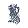
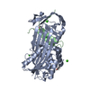
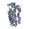
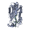
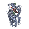
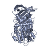
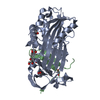
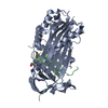
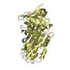
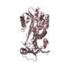
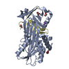
| Title | CLEAVED ANTICHYMOTRYPSIN A349R | ||||||
 Components Components | (ANTICHYMOTRYPSIN) x 2 | ||||||
 Keywords Keywords |  SERPIN / SERINE PROTEASE INHIBITOR / ANTICHYMOTRYPSIN SERPIN / SERINE PROTEASE INHIBITOR / ANTICHYMOTRYPSIN | ||||||
| Function / homology |  Function and homology information Function and homology informationmaintenance of gastrointestinal epithelium / regulation of lipid metabolic process / platelet alpha granule lumen / acute-phase response / serine-type endopeptidase inhibitor activity / azurophil granule lumen / Platelet degranulation / collagen-containing extracellular matrix / blood microparticle / secretory granule lumen ...maintenance of gastrointestinal epithelium / regulation of lipid metabolic process / platelet alpha granule lumen / acute-phase response / serine-type endopeptidase inhibitor activity / azurophil granule lumen / Platelet degranulation / collagen-containing extracellular matrix / blood microparticle / secretory granule lumen / inflammatory response / Neutrophil degranulation / DNA binding / extracellular space / extracellular exosome / extracellular region / nucleusSimilarity search - Function | ||||||
| Biological species |  Homo sapiens (human) Homo sapiens (human) | ||||||
| Method | X-RAY DIFFRACTION / DIFFERENCE FOURIER / Resolution: 2.1 Å | ||||||
 Authors Authors | Lukacs, C.M. / Christianson, D.W. | ||||||
 Citation Citation | Journal: Biochemistry / Year: 1998 Title: Engineering an anion-binding cavity in antichymotrypsin modulates the "spring-loaded" serpin-protease interaction. Authors: Lukacs, C.M. / Rubin, H. / Christianson, D.W. #1: Journal: Nat.Struct.Biol. / Year: 1996Title: Arginine Substitutions in the Hinge Region of Antichymotrypsin Affect Serpin Beta-Sheet Rearrangement Authors: Lukacs, C.M. / Zhong, J.Q. / Plotnick, M.I. / Rubin, H. / Cooperman, B.S. / Christianson, D.W. | ||||||
| History |
|
- Structure visualization
| Structure viewer | Molecule: MolmilJmol/JSmol |
|---|
- Downloads & links
-Download
| PDBx/mmCIF format | 1as4.cif.gz | 86.3 KB | Display | PDBx/mmCIF format |
|---|---|---|---|---|
| PDB format | pdb1as4.ent.gz | 64.9 KB | Display | PDB format |
| PDBx/mmJSON format | 1as4.json.gz | Tree view | PDBx/mmJSON format | |
| Others |  Other downloads Other downloads |
-Validation report
| Arichive directory | https://data.pdbj.org/pub/pdb/validation_reports/as/1as4ftp://data.pdbj.org/pub/pdb/validation_reports/as/1as4 | HTTPS FTP |
|---|
-Related structure data


-Links
 PDBj
PDBj
- Assembly
| Deposited unit | 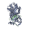
| ||||||||
|---|---|---|---|---|---|---|---|---|---|
| 1 |
| ||||||||
| Unit cell |
|
-Components
| #1: Protein | Mass: 38612.148 Da / Num. of mol.: 1 Fragment: CHAIN A CONTAINS RESIDUES 20 - 358, CHAIN B CONTAINS RESIDUES 359 - 393 Mutation: A349R Source method: isolated from a genetically manipulated source Details: CLEAVED ANTICHYMOTRYPSIN / Source: (gene. exp.) Homo sapiens (human) / Gene: ACT / Plasmid: PZMS / Gene (production host): ACT / Production host:  Escherichia coli (E. coli) / References: UniProt: P01011 Escherichia coli (E. coli) / References: UniProt: P01011 |
|---|---|
| #2: Protein/peptide | Mass: 4259.000 Da / Num. of mol.: 1 Fragment: CHAIN A CONTAINS RESIDUES 20 - 358, CHAIN B CONTAINS RESIDUES 359 - 393 Mutation: A349R Source method: isolated from a genetically manipulated source Details: CLEAVED ANTICHYMOTRYPSIN / Source: (gene. exp.) Homo sapiens (human) / Gene: ACT / Plasmid: PZMS / Gene (production host): ACT / Production host: Escherichia coli (E. coli) / References: UniProt: P01011 |
| #3: Chemical | ChemComp-ACT / Acetate  Mass: 59.044 Da / Num. of mol.: 1 / Source method: obtained synthetically / Formula: C2H3O2 Mass: 59.044 Da / Num. of mol.: 1 / Source method: obtained synthetically / Formula: C2H3O2 |
| #4: Water | ChemComp-HOH / Water Mass: 18.015 Da / Num. of mol.: 57 / Source method: isolated from a natural source / Formula: H2O Mass: 18.015 Da / Num. of mol.: 57 / Source method: isolated from a natural source / Formula: H2O |
| Compound details | RESIDUE 349 IS AN ALA -> ARG MUTATION. THE ARGININE IS BURIED WITH AN ACETATE COUNTERION |
-Experimental details
-Experiment
| Experiment | Method: X-RAY DIFFRACTION / Number of used crystals: 1 |
|---|
- Sample preparation
| Crystal | Density Matthews: 2.65 Å3/Da / Density % sol: 53 % / Description: COORDINATES FROM 2CAA USED FOR DIFF. FOUR. | ||||||||||||||||||||||||||||||||||||||||
|---|---|---|---|---|---|---|---|---|---|---|---|---|---|---|---|---|---|---|---|---|---|---|---|---|---|---|---|---|---|---|---|---|---|---|---|---|---|---|---|---|---|
| Crystal grow | pH: 5.6 Details: 14% PEG MONOMETHYLETHER 5000, 0.2 M MAGNESIUM ACETATE 0.1 M SODIUM ACETATE PH 5.6 PROTEIN AT 3 MG/ML | ||||||||||||||||||||||||||||||||||||||||
| Crystal grow | *PLUS Method: vapor diffusion, hanging drop | ||||||||||||||||||||||||||||||||||||||||
| Components of the solutions | *PLUS
|
-Data collection
| Diffraction | Mean temperature: 298 K |
|---|---|
| Diffraction source | Source: ROTATING ANODE / Type: RIGAKU RUH2R / Wavelength: 1.5418 |
| Detector | Type: RIGAKU RAXIS II / Detector: IMAGE PLATE / Date: Jan 1, 1997 / Details: MIRRORS |
| Radiation | Monochromatic (M) / Laue (L): M / Scattering type: x-ray |
| Radiation wavelength | Wavelength: 1.5418 Å / Relative weight: 1 |
| Reflection | Resolution: 2.1→20 Å / Num. obs: 26288 / % possible obs: 98.7 % / Redundancy: 3.6 % / Biso Wilson estimate: 24.76 Å2 / Rmerge(I) obs: 0.066 / Net I/σ(I): 8.9 |
| Reflection shell | Resolution: 2.1→2.15 Å / Redundancy: 2.9 % / Rmerge(I) obs: 0.235 / Mean I/σ(I) obs: 2.6 / % possible all: 93.8 |
| Reflection | *PLUS Num. measured all: 95680 |
- Processing
| Software |
| ||||||||||||||||||||||||||||||||||||||||||||||||||||||||||||
|---|---|---|---|---|---|---|---|---|---|---|---|---|---|---|---|---|---|---|---|---|---|---|---|---|---|---|---|---|---|---|---|---|---|---|---|---|---|---|---|---|---|---|---|---|---|---|---|---|---|---|---|---|---|---|---|---|---|---|---|---|---|
| Refinement | Method to determine structure: DIFFERENCE FOURIER / Resolution: 2.1→20 Å / Rfactor Rfree error: 0.0075 / Data cutoff high absF: 100000 / Data cutoff low absF: 0.1 / Cross valid method: THROUGHOUT / σ(F): 1 Details: RESIDUES GLN 105 - GLU 109 ARE IN VERY POOR DENSITY, AND THEIR CONFORMATIONS SHOULD NOT BE CONSIDERED AS ACTUAL.
| ||||||||||||||||||||||||||||||||||||||||||||||||||||||||||||
| Displacement parameters | Biso mean: 30.3 Å2 | ||||||||||||||||||||||||||||||||||||||||||||||||||||||||||||
| Refine analyze | Luzzati d res low obs: 20 Å | ||||||||||||||||||||||||||||||||||||||||||||||||||||||||||||
| Refinement step | Cycle: LAST / Resolution: 2.1→20 Å
| ||||||||||||||||||||||||||||||||||||||||||||||||||||||||||||
| Refine LS restraints |
| ||||||||||||||||||||||||||||||||||||||||||||||||||||||||||||
| LS refinement shell | Resolution: 2.1→2.2 Å / Rfactor Rfree error: 0.029 / Total num. of bins used: 8
| ||||||||||||||||||||||||||||||||||||||||||||||||||||||||||||
| Xplor file |
| ||||||||||||||||||||||||||||||||||||||||||||||||||||||||||||
| Software | *PLUS Name: X-PLOR / Version: 3.1 / Classification: refinement | ||||||||||||||||||||||||||||||||||||||||||||||||||||||||||||
| Refinement | *PLUS Rfactor Rfree: 0.24 | ||||||||||||||||||||||||||||||||||||||||||||||||||||||||||||
| Solvent computation | *PLUS | ||||||||||||||||||||||||||||||||||||||||||||||||||||||||||||
| Displacement parameters | *PLUS | ||||||||||||||||||||||||||||||||||||||||||||||||||||||||||||
| Refine LS restraints | *PLUS
|
