 Movie
Movie Controller

[English] 日本語
 Yorodumi
- PDB-1apl: CRYSTAL STRUCTURE OF A MAT-ALPHA2 HOMEODOMAIN-OPERATOR COMPLEX SU... -
+ Open data

- Basic information
| Entry | Database: PDB / ID: 1apl | ||||||
|---|---|---|---|---|---|---|---|
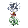
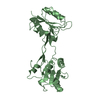
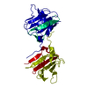
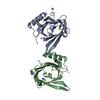
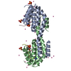
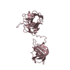
| Title | CRYSTAL STRUCTURE OF A MAT-ALPHA2 HOMEODOMAIN-OPERATOR COMPLEX SUGGESTS A GENERAL MODEL FOR HOMEODOMAIN-DNA INTERACTIONS | ||||||
 Components Components |
| ||||||
 Keywords Keywords | DNA BINDING PROTEIN/DNA /  PROTEIN-DNA COMPLEX / DOUBLE HELIX / DNA BINDING PROTEIN-DNA COMPLEX PROTEIN-DNA COMPLEX / DOUBLE HELIX / DNA BINDING PROTEIN-DNA COMPLEX | ||||||
| Function / homology |  Function and homology information Function and homology information: / regulation of mating-type specific transcription, DNA-templated / RNA polymerase II transcription repressor complex / DNA binding, bending / DNA-binding transcription repressor activity, RNA polymerase II-specific / sequence-specific DNA binding / DNA-binding transcription factor activity, RNA polymerase II-specific / RNA polymerase II cis-regulatory region sequence-specific DNA binding / regulation of transcription by RNA polymerase II / negative regulation of transcription by RNA polymerase II / nucleusSimilarity search - Function | ||||||
| Biological species |  Saccharomyces cerevisiae (brewer's yeast) Saccharomyces cerevisiae (brewer's yeast) | ||||||
| Method | X-RAY DIFFRACTION / Resolution: 2.7 Å | ||||||
 Authors Authors | Wolberger, C. / Vershon, A.K. / Liu, B. / Johnson, A.D. / Pabo, C.O. | ||||||
 Citation Citation | Journal: Cell(Cambridge,Mass.) / Year: 1991 Title: Crystal structure of a MAT alpha 2 homeodomain-operator complex suggests a general model for homeodomain-DNA interactions. Authors: Wolberger, C. / Vershon, A.K. / Liu, B. / Johnson, A.D. / Pabo, C.O. #1: Journal: J.Mol.Biol. / Year: 1991Title: Crystallization and Preliminary X-Ray Diffraction Studies of a MAT-Alpha2-DNA Complex Authors: Wolberger, C. / Vershon, A.K. / Johnson, A.D. / Pabo, C.O. | ||||||
| History |
|
- Structure visualization
| Structure viewer | Molecule: MolmilJmol/JSmol |
|---|
- Downloads & links
-Download
| PDBx/mmCIF format | 1apl.cif.gz | 63.2 KB | Display | PDBx/mmCIF format |
|---|---|---|---|---|
| PDB format | pdb1apl.ent.gz | 41.3 KB | Display | PDB format |
| PDBx/mmJSON format | 1apl.json.gz | Tree view | PDBx/mmJSON format | |
| Others |  Other downloads Other downloads |
-Validation report
| Arichive directory | https://data.pdbj.org/pub/pdb/validation_reports/ap/1aplftp://data.pdbj.org/pub/pdb/validation_reports/ap/1apl | HTTPS FTP |
|---|
-Related structure data
| Similar structure data |
|---|


-Links
 PDBj
PDBj


- Assembly
| Deposited unit | 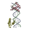
| ||||||||
|---|---|---|---|---|---|---|---|---|---|
| 1 |
| ||||||||
| Unit cell |
|
-Components
| #1: DNA chain | Mass: 6381.160 Da / Num. of mol.: 1 / Source method: obtained synthetically |
|---|---|
| #2: DNA chain | Mass: 6501.232 Da / Num. of mol.: 1 / Source method: obtained synthetically |
| #3: Protein | Mass: 9773.306 Da / Num. of mol.: 2 Source method: isolated from a genetically manipulated source Source: (gene. exp.) Saccharomyces cerevisiae (brewer's yeast)Gene: MAT ALPHA2 RES. 128-210 / Plasmid: PAV56 / Gene (production host): MAT ALPHA2 RES. 128-210 / Production host:  Escherichia coli (E. coli) / References: UniProt: Q6B2C0, UniProt: P0CY08*PLUS Escherichia coli (E. coli) / References: UniProt: Q6B2C0, UniProt: P0CY08*PLUS |
-Experimental details
-Experiment
| Experiment | Method: X-RAY DIFFRACTION |
|---|
- Sample preparation
| Crystal | Density Matthews: 2.52 Å3/Da / Density % sol: 51 % | ||||||||||||||||||||||||||||||||||||||||||
|---|---|---|---|---|---|---|---|---|---|---|---|---|---|---|---|---|---|---|---|---|---|---|---|---|---|---|---|---|---|---|---|---|---|---|---|---|---|---|---|---|---|---|---|
| Crystal grow | Method: vapor diffusion, hanging drop / pH: 7.5 / Details: pH 7.50, VAPOR DIFFUSION, HANGING DROP / Temp details: ROOM TEMPERATURE | ||||||||||||||||||||||||||||||||||||||||||
| Components of the solutions |
| ||||||||||||||||||||||||||||||||||||||||||
| Crystal | *PLUS | ||||||||||||||||||||||||||||||||||||||||||
| Crystal grow | *PLUS pH: 7.5 / Method: vapor diffusion, hanging drop / Details: referred to J.Mol.Biol. 217.11-13 1991 | ||||||||||||||||||||||||||||||||||||||||||
| Components of the solutions | *PLUS
|
-Data collection
| Diffraction source | Source: ROTATING ANODE / Type: RIGAKU RU200 |
|---|---|
| Detector | Type: SIEMENS / Detector: AREA DETECTOR |
| Radiation | Protocol: SINGLE WAVELENGTH / Monochromatic (M) / Laue (L): M / Scattering type: x-ray |
| Radiation wavelength | Relative weight: 1 |
| Reflection | Highest resolution: 2.9 Å / Num. obs: 7334 / % possible obs: 90 % / Observed criterion σ(I): 1 |
| Reflection | *PLUS Highest resolution: 2.65 Å / Num. obs: 7907 / Num. measured all: 18265 / Rmerge(I) obs: 0.075 |
- Processing
| Software | Name: X-PLOR / Classification: refinement | ||||||||||||||||||||||||||||||||||||||||||||||||||||||||||||
|---|---|---|---|---|---|---|---|---|---|---|---|---|---|---|---|---|---|---|---|---|---|---|---|---|---|---|---|---|---|---|---|---|---|---|---|---|---|---|---|---|---|---|---|---|---|---|---|---|---|---|---|---|---|---|---|---|---|---|---|---|---|
| Refinement | Resolution: 2.7→8 Å / σ(F): 1 /
| ||||||||||||||||||||||||||||||||||||||||||||||||||||||||||||
| Refinement step | Cycle: LAST / Resolution: 2.7→8 Å
| ||||||||||||||||||||||||||||||||||||||||||||||||||||||||||||
| Refine LS restraints |
| ||||||||||||||||||||||||||||||||||||||||||||||||||||||||||||
| Refinement | *PLUS Highest resolution: 2.7 Å / Lowest resolution: 8 Å / σ(I): 1 / Rfactor obs: 0.226 | ||||||||||||||||||||||||||||||||||||||||||||||||||||||||||||
| Solvent computation | *PLUS | ||||||||||||||||||||||||||||||||||||||||||||||||||||||||||||
| Displacement parameters | *PLUS | ||||||||||||||||||||||||||||||||||||||||||||||||||||||||||||
| Refine LS restraints | *PLUS
|
