 Movie
Movie Controller


+ Open data

- Basic information
| Entry | Database: PDB / ID: 1aow | ||||||
|---|---|---|---|---|---|---|---|
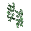
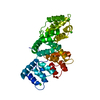
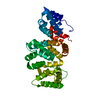
| Title | ANNEXIN IV | ||||||
 Components Components | ANNEXIN IV | ||||||
 Keywords Keywords | CALCIUM/PHOSPHOLIPID-BINDING PROTEIN / 32.5KD CALELECTRIN / ENDONEXIN I / LIPOCORTIN IV / CHROMOBINDIN IV / PROTEIN II / CALCIUM-PHOSPHOLIPID-BINDING PROTEIN complex | ||||||
| Function / homology |  Function and homology informationchondroitin sulfate binding / zymogen granule membrane / chromaffin granule membrane / negative regulation of interleukin-8 production / calcium-dependent phospholipid binding / NF-kappaB binding / Notch signaling pathway / epithelial cell differentiation / calcium-dependent protein binding / heparin binding ...chondroitin sulfate binding / zymogen granule membrane / chromaffin granule membrane / negative regulation of interleukin-8 production / calcium-dependent phospholipid binding / NF-kappaB binding / Notch signaling pathway / epithelial cell differentiation / calcium-dependent protein binding / heparin binding / carbohydrate binding / nuclear membrane / apical plasma membrane / calcium ion binding / regulation of transcription by RNA polymerase II / perinuclear region of cytoplasm / cell surface / membrane / identical protein binding / nucleus / cytosol / cytoplasm Function and homology informationchondroitin sulfate binding / zymogen granule membrane / chromaffin granule membrane / negative regulation of interleukin-8 production / calcium-dependent phospholipid binding / NF-kappaB binding / Notch signaling pathway / epithelial cell differentiation / calcium-dependent protein binding / heparin binding ...chondroitin sulfate binding / zymogen granule membrane / chromaffin granule membrane / negative regulation of interleukin-8 production / calcium-dependent phospholipid binding / NF-kappaB binding / Notch signaling pathway / epithelial cell differentiation / calcium-dependent protein binding / heparin binding / carbohydrate binding / nuclear membrane / apical plasma membrane / calcium ion binding / regulation of transcription by RNA polymerase II / perinuclear region of cytoplasm / cell surface / membrane / identical protein binding / nucleus / cytosol / cytoplasmSimilarity search - Function | ||||||
| Biological species |  Bos taurus (cattle) Bos taurus (cattle) | ||||||
| Method | X-RAY DIFFRACTION / MOLECULAR REPLACEMENT / Resolution: 3 Å | ||||||
 Authors Authors | Zanotti, G. / Malpeli, G. / Gliubich, F. / Folli, C. / Stoppini, M. / Olivi, L. / Savoia, A. / Berni, R. | ||||||
 Citation Citation | Journal: Biochem.J. / Year: 1998 Title: Structure of the trigonal crystal form of bovine annexin IV. Authors: Zanotti, G. / Malpeli, G. / Gliubich, F. / Folli, C. / Stoppini, M. / Olivi, L. / Savoia, A. / Berni, R. | ||||||
| History |
|
- Structure visualization
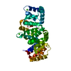
| Structure viewer | Molecule: MolmilJmol/JSmol |
|---|
- Downloads & links
-Download
| PDBx/mmCIF format | 1aow.cif.gz | 67.1 KB | Display | PDBx/mmCIF format |
|---|---|---|---|---|
| PDB format | pdb1aow.ent.gz | 53.3 KB | Display | PDB format |
| PDBx/mmJSON format | 1aow.json.gz | Tree view | PDBx/mmJSON format | |
| Others |  Other downloads Other downloads |
-Validation report
| Arichive directory | https://data.pdbj.org/pub/pdb/validation_reports/ao/1aowftp://data.pdbj.org/pub/pdb/validation_reports/ao/1aow | HTTPS FTP |
|---|
-Related structure data
| Related structure data |  1annS S: Starting model for refinement |
|---|---|
| Similar structure data |


-Links
 PDBj
PDBj- Assembly
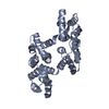
| Deposited unit | 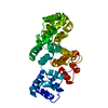
| ||||||||
|---|---|---|---|---|---|---|---|---|---|
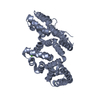
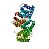
| 1 |
| ||||||||
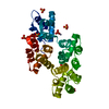
| Unit cell |
|
-Components
| #1: Protein | / 32.5 KD CALELECTRIN / ENDONEXIN I Mass: 35005.648 Da / Num. of mol.: 1 / Source method: isolated from a natural source / Source: (natural) Bos taurus (cattle) / Organ: KIDNEY / References: UniProt: P13214 |
|---|
-Experimental details
-Experiment
| Experiment | Method: X-RAY DIFFRACTION / Number of used crystals: 1 |
|---|
- Sample preparation
| Crystal | Density Matthews: 3.37 Å3/Da / Density % sol: 63 % | |||||||||||||||||||||||||||||||||||
|---|---|---|---|---|---|---|---|---|---|---|---|---|---|---|---|---|---|---|---|---|---|---|---|---|---|---|---|---|---|---|---|---|---|---|---|---|
| Crystal grow | Method: vapor diffusion, sitting drop / pH: 5 Details: SITTING DROP, FROM A PROTEIN SOLUTION 10 MG/ML, IN THE PRESENCE OF 1.4 M AMMONIUM SULFATE, 10 MM BETA-MERCAPTOETHANOL, PH=5.0, vapor diffusion - sitting drop | |||||||||||||||||||||||||||||||||||
| Crystal grow | *PLUS Method: vapor diffusion, sitting drop | |||||||||||||||||||||||||||||||||||
| Components of the solutions | *PLUS
|
-Data collection
| Diffraction | Mean temperature: 100 K |
|---|---|
| Diffraction source | Wavelength: 1.3 |
| Detector | Type: MARRESEARCH / Detector: IMAGE PLATE AREA DETECTOR / Date: Feb 1, 1997 / Details: MIRRORS |
| Radiation | Monochromator: SI(111) / Monochromatic (M) / Laue (L): M / Scattering type: x-ray |
| Radiation wavelength | Wavelength: 1.3 Å / Relative weight: 1 |
| Reflection | Resolution: 3→15 Å / Num. obs: 8114 / % possible obs: 95 % / Redundancy: 5.6 % / Rmerge(I) obs: 0.049 / Rsym value: 0.049 / Net I/σ(I): 36 |
| Reflection shell | Resolution: 2.99→3.12 Å / Redundancy: 3 % / Rmerge(I) obs: 0.07 / Mean I/σ(I) obs: 10 / Rsym value: 0.07 / % possible all: 75.3 |
| Reflection | *PLUS Highest resolution: 2.99 Å / Lowest resolution: 50 Å / % possible obs: 93 % |
| Reflection shell | *PLUS Lowest resolution: 3.5 Å / % possible obs: 87 % |
- Processing
| Software |
| ||||||||||||||||||||||||||||||||||||||||||||||||||||||||||||
|---|---|---|---|---|---|---|---|---|---|---|---|---|---|---|---|---|---|---|---|---|---|---|---|---|---|---|---|---|---|---|---|---|---|---|---|---|---|---|---|---|---|---|---|---|---|---|---|---|---|---|---|---|---|---|---|---|---|---|---|---|---|
| Refinement | Method to determine structure: MOLECULAR REPLACEMENT Starting model: PDB ENTRY 1ANN Resolution: 3→15 Å / Data cutoff high absF: 10000000 / Data cutoff low absF: 0 / Cross valid method: THROUGHOUT / σ(F): 0
| ||||||||||||||||||||||||||||||||||||||||||||||||||||||||||||
| Displacement parameters | Biso mean: 20.5 Å2 | ||||||||||||||||||||||||||||||||||||||||||||||||||||||||||||
| Refinement step | Cycle: LAST / Resolution: 3→15 Å
| ||||||||||||||||||||||||||||||||||||||||||||||||||||||||||||
| Refine LS restraints |
| ||||||||||||||||||||||||||||||||||||||||||||||||||||||||||||
| Xplor file |
| ||||||||||||||||||||||||||||||||||||||||||||||||||||||||||||
| Software | *PLUS Name: X-PLOR / Version: 3.1 / Classification: refinement | ||||||||||||||||||||||||||||||||||||||||||||||||||||||||||||
| Refinement | *PLUS Highest resolution: 2.99 Å / Lowest resolution: 7 Å / Num. reflection obs: 6717 / Num. reflection Rfree: 800 | ||||||||||||||||||||||||||||||||||||||||||||||||||||||||||||
| Solvent computation | *PLUS | ||||||||||||||||||||||||||||||||||||||||||||||||||||||||||||
| Displacement parameters | *PLUS |
