 Movie
Movie Controller


+ Open data

- Basic information
| Entry | Database: PDB / ID: 1aok | ||||||
|---|---|---|---|---|---|---|---|
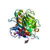
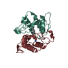
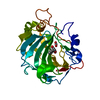
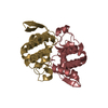
| Title | VIPOXIN COMPLEX | ||||||
 Components Components | (VIPOXIN COMPLEX) x 2 | ||||||
 Keywords Keywords |  HYDROLASE / PHOSPHOLIPASE / VIPOXIN / PLA2-ACTIVITY / SNAKE-VENOM HYDROLASE / PHOSPHOLIPASE / VIPOXIN / PLA2-ACTIVITY / SNAKE-VENOM | ||||||
| Function / homology |  Function and homology informationphospholipase A2 / phospholipase A2 activity / arachidonic acid secretion / phospholipid metabolic process / lipid catabolic process / toxin activity / killing of cells of another organism / calcium ion binding / extracellular region Function and homology informationphospholipase A2 / phospholipase A2 activity / arachidonic acid secretion / phospholipid metabolic process / lipid catabolic process / toxin activity / killing of cells of another organism / calcium ion binding / extracellular regionSimilarity search - Function | ||||||
| Biological species |  Vipera ammodytes meridionalis (snake) Vipera ammodytes meridionalis (snake) | ||||||
| Method | X-RAY DIFFRACTION / SYNCHROTRON / MOLECULAR REPLACEMENT / Resolution: 2 Å | ||||||
 Authors Authors | Perbandt, M. / Wilson, J.C. / Eschenburg, S. / Betzel, C. | ||||||
 Citation Citation | Journal: FEBS Lett. / Year: 1997 Title: Crystal structure of vipoxin at 2.0 A: an example of regulation of a toxic function generated by molecular evolution. Authors: Perbandt, M. / Wilson, J.C. / Eschenburg, S. / Mancheva, I. / Aleksiev, B. / Genov, N. / Willingmann, P. / Weber, W. / Singh, T.P. / Betzel, C. | ||||||
| History |
|
- Structure visualization
| Structure viewer | Molecule: MolmilJmol/JSmol |
|---|
- Downloads & links
-Download
| PDBx/mmCIF format | 1aok.cif.gz | 61.7 KB | Display | PDBx/mmCIF format |
|---|---|---|---|---|
| PDB format | pdb1aok.ent.gz | 48.4 KB | Display | PDB format |
| PDBx/mmJSON format | 1aok.json.gz | Tree view | PDBx/mmJSON format | |
| Others |  Other downloads Other downloads |
-Validation report
| Arichive directory | https://data.pdbj.org/pub/pdb/validation_reports/ao/1aokftp://data.pdbj.org/pub/pdb/validation_reports/ao/1aok | HTTPS FTP |
|---|
-Related structure data
| Related structure data |  1pp2S S: Starting model for refinement |
|---|---|
| Similar structure data |


-Links
 PDBj
PDBj

- Assembly
| Deposited unit | 
| ||||||||
|---|---|---|---|---|---|---|---|---|---|
| 1 |
| ||||||||
| Unit cell |
|
-Components
| #1: Protein | Mass: 13661.036 Da / Num. of mol.: 1 / Source method: isolated from a natural source / Source: (natural) Vipera ammodytes meridionalis (snake) / Secretion: VENOM / Species: Vipera ammodytes / Strain: meridionalis / References: UniProt: P04084, phospholipase A2 |
|---|---|
| #2: Protein | Mass: 13835.843 Da / Num. of mol.: 1 / Source method: isolated from a natural source / Source: (natural) Vipera ammodytes meridionalis (snake) / Secretion: VENOM / Species: Vipera ammodytes / Strain: meridionalis / References: UniProt: P14420, phospholipase A2 |
| #3: Chemical | ChemComp-ACT / Acetate  Mass: 59.044 Da / Num. of mol.: 1 / Source method: obtained synthetically / Formula: C2H3O2 Mass: 59.044 Da / Num. of mol.: 1 / Source method: obtained synthetically / Formula: C2H3O2 |
| #4: Water | ChemComp-HOH / Water Mass: 18.015 Da / Num. of mol.: 291 / Source method: isolated from a natural source / Formula: H2O Mass: 18.015 Da / Num. of mol.: 291 / Source method: isolated from a natural source / Formula: H2O |
| Compound details | IN VIPOXIN ONE SUBUNIT IS ROTATED BY ABOUT 180 DEGREE TO GIVE A PSEUDO MIRROR SYMMETRY AND THE ...IN VIPOXIN ONE SUBUNIT IS ROTATED BY ABOUT 180 DEGREE TO GIVE A PSEUDO MIRROR SYMMETRY AND THE COMPLEX IS STABILIZED |
-Experimental details
-Experiment
| Experiment | Method: X-RAY DIFFRACTION / Number of used crystals: 1 |
|---|
- Sample preparation
| Crystal | Density Matthews: 2 Å3/Da / Density % sol: 36.88 % | ||||||||||||||||||||||||||||||||||||
|---|---|---|---|---|---|---|---|---|---|---|---|---|---|---|---|---|---|---|---|---|---|---|---|---|---|---|---|---|---|---|---|---|---|---|---|---|---|
| Crystal grow | pH: 4.8 / Details: pH 4.8 | ||||||||||||||||||||||||||||||||||||
| Crystal grow | *PLUS Temperature: 20 ℃ / Method: vapor diffusion, sitting drop | ||||||||||||||||||||||||||||||||||||
| Components of the solutions | *PLUS
|
-Data collection
| Diffraction | Mean temperature: 100 K |
|---|---|
| Diffraction source | Source: SYNCHROTRON / Site: EMBL/DESY, HAMBURG  / Beamline: BW7B / Wavelength: 0.92 / Beamline: BW7B / Wavelength: 0.92 |
| Detector | Type: MARRESEARCH / Detector: IMAGE PLATE / Date: Sep 15, 1995 / Details: COLLIMATOR |
| Radiation | Monochromatic (M) / Laue (L): M / Scattering type: x-ray |
| Radiation wavelength | Wavelength: 0.92 Å / Relative weight: 1 |
| Reflection | Resolution: 2→20 Å / Num. obs: 15070 / % possible obs: 100 % / Observed criterion σ(I): 2 / Rsym value: 0.086 |
| Reflection shell | Resolution: 2→2.03 Å / Rsym value: 0.274 / % possible all: 100 |
| Reflection | *PLUS Rmerge(I) obs: 0.086 |
| Reflection shell | *PLUS % possible obs: 100 % / Rmerge(I) obs: 0.274 |
- Processing
| Software |
| |||||||||||||||||||||||||||||||||||||||||||||||||||||||||||||||
|---|---|---|---|---|---|---|---|---|---|---|---|---|---|---|---|---|---|---|---|---|---|---|---|---|---|---|---|---|---|---|---|---|---|---|---|---|---|---|---|---|---|---|---|---|---|---|---|---|---|---|---|---|---|---|---|---|---|---|---|---|---|---|---|---|
| Refinement | Method to determine structure: MOLECULAR REPLACEMENT Starting model: PDB ENTRY 1PP2 Resolution: 2→20 Å /
| |||||||||||||||||||||||||||||||||||||||||||||||||||||||||||||||
| Refinement step | Cycle: LAST / Resolution: 2→20 Å
| |||||||||||||||||||||||||||||||||||||||||||||||||||||||||||||||
| Refine LS restraints |
| |||||||||||||||||||||||||||||||||||||||||||||||||||||||||||||||
| Software | *PLUS Name: REFMAC / Classification: refinement | |||||||||||||||||||||||||||||||||||||||||||||||||||||||||||||||
| Refinement | *PLUS Rfactor obs: 0.16 | |||||||||||||||||||||||||||||||||||||||||||||||||||||||||||||||
| Solvent computation | *PLUS | |||||||||||||||||||||||||||||||||||||||||||||||||||||||||||||||
| Displacement parameters | *PLUS |
