 Movie
Movie Controller

[English] 日本語
 Yorodumi
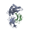
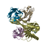
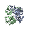
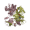
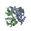
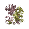
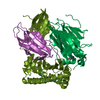
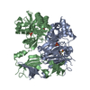
- PDB-1akj: COMPLEX OF THE HUMAN MHC CLASS I GLYCOPROTEIN HLA-A2 AND THE T CE... -
+ Open data

- Basic information
| Entry | Database: PDB / ID: 1akj | ||||||
|---|---|---|---|---|---|---|---|
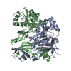
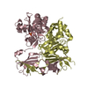
| Title | COMPLEX OF THE HUMAN MHC CLASS I GLYCOPROTEIN HLA-A2 AND THE T CELL CORECEPTOR CD8 | ||||||
 Components Components |
| ||||||
 Keywords Keywords | COMPLEX (MHC I/PEPTIDE/CD8) /  T-CELL / GLYCOPROTEIN / COMPLEX / IMMUNOLOGY / COMPLEX (MHC I-PEPTIDE-CD8) / COMPLEX (MHC I-PEPTIDE-CD8) complex T-CELL / GLYCOPROTEIN / COMPLEX / IMMUNOLOGY / COMPLEX (MHC I-PEPTIDE-CD8) / COMPLEX (MHC I-PEPTIDE-CD8) complex | ||||||
| Function / homology |  Function and homology information Function and homology informationcytotoxic T cell differentiation / MHC class I protein complex binding / T cell mediated immunity / T cell receptor complex / plasma membrane raft / T cell mediated cytotoxicity directed against tumor cell target / antigen processing and presentation of endogenous peptide antigen via MHC class I via ER pathway, TAP-dependent / positive regulation of memory T cell activation / TAP complex binding / antigen processing and presentation of exogenous peptide antigen via MHC class I ...cytotoxic T cell differentiation / MHC class I protein complex binding / T cell mediated immunity / T cell receptor complex / plasma membrane raft / T cell mediated cytotoxicity directed against tumor cell target / antigen processing and presentation of endogenous peptide antigen via MHC class I via ER pathway, TAP-dependent / positive regulation of memory T cell activation / TAP complex binding / antigen processing and presentation of exogenous peptide antigen via MHC class I / Golgi medial cisterna / positive regulation of CD8-positive, alpha-beta T cell activation / CD8-positive, alpha-beta T cell activation / positive regulation of CD8-positive, alpha-beta T cell proliferation / CD8 receptor binding / antigen processing and presentation / MHC class I protein binding / endoplasmic reticulum exit site / coreceptor activity / beta-2-microglobulin binding / TAP binding / protection from natural killer cell mediated cytotoxicity / antigen processing and presentation of endogenous peptide antigen via MHC class I via ER pathway, TAP-independent / antigen processing and presentation of endogenous peptide antigen via MHC class Ib / detection of bacterium / T cell activation / T cell receptor binding / positive regulation of ferrous iron binding / positive regulation of transferrin receptor binding / early endosome lumen / positive regulation of receptor binding / Nef mediated downregulation of MHC class I complex cell surface expression / DAP12 interactions / negative regulation of receptor binding / lumenal side of endoplasmic reticulum membrane / Endosomal/Vacuolar pathway / Antigen Presentation: Folding, assembly and peptide loading of class I MHC / antigen processing and presentation of exogenous protein antigen via MHC class Ib, TAP-dependent / cellular response to iron(III) ion / negative regulation of forebrain neuron differentiation / ER to Golgi transport vesicle membrane / response to molecule of bacterial origin / regulation of erythrocyte differentiation / regulation of iron ion transport / MHC class I peptide loading complex / HFE-transferrin receptor complex / T cell mediated cytotoxicity / cellular response to iron ion / antigen processing and presentation of endogenous peptide antigen via MHC class I / positive regulation of T cell cytokine production / MHC class I protein complex / multicellular organismal-level iron ion homeostasis / positive regulation of T cell mediated cytotoxicity / peptide antigen assembly with MHC class II protein complex / negative regulation of neurogenesis / positive regulation of receptor-mediated endocytosis / MHC class II protein complex / cellular response to nicotine / recycling endosome membrane / specific granule lumen / phagocytic vesicle membrane / peptide antigen binding / cell surface receptor protein tyrosine kinase signaling pathway / positive regulation of cellular senescence / antigen processing and presentation of exogenous peptide antigen via MHC class II / Immunoregulatory interactions between a Lymphoid and a non-Lymphoid cell / Interferon gamma signaling / positive regulation of immune response / negative regulation of epithelial cell proliferation / Modulation by Mtb of host immune system / positive regulation of T cell activation / Interferon alpha/beta signaling / positive regulation of type II interferon production / sensory perception of smell / negative regulation of neuron projection development / E3 ubiquitin ligases ubiquitinate target proteins / tertiary granule lumen / DAP12 signaling / MHC class II protein complex binding / late endosome membrane / T cell differentiation in thymus / positive regulation of protein binding / ER-Phagosome pathway / antibacterial humoral response / iron ion transport / T cell receptor signaling pathway / protein refolding / early endosome membrane / protein homotetramerization / intracellular iron ion homeostasis / amyloid fibril formation / adaptive immune response / learning or memory / cell surface receptor signaling pathway / receptor complex / defense response to Gram-positive bacterium / immune response / Amyloid fiber formation / lysosomal membrane / endoplasmic reticulum lumenSimilarity search - Function | ||||||
| Biological species |  Homo sapiens (human) Homo sapiens (human)  Human immunodeficiency virus Human immunodeficiency virus | ||||||
| Method | X-RAY DIFFRACTION / MOLECULAR REPLACEMENT / Resolution: 2.65 Å | ||||||
 Authors Authors | Tormo, J. / Stuart, D.I. / Jones, E.Y. | ||||||
 Citation Citation | Journal: Nature / Year: 1997 Title: Crystal structure of the complex between human CD8alpha(alpha) and HLA-A2. Authors: Gao, G.F. / Tormo, J. / Gerth, U.C. / Wyer, J.R. / McMichael, A.J. / Stuart, D.I. / Bell, J.I. / Jones, E.Y. / Jakobsen, B.K. | ||||||
| History |
|
- Structure visualization
| Structure viewer | Molecule: MolmilJmol/JSmol |
|---|
- Downloads & links
-Download
| PDBx/mmCIF format | 1akj.cif.gz | 130.9 KB | Display | PDBx/mmCIF format |
|---|---|---|---|---|
| PDB format | pdb1akj.ent.gz | 105.2 KB | Display | PDB format |
| PDBx/mmJSON format | 1akj.json.gz | Tree view | PDBx/mmJSON format | |
| Others |  Other downloads Other downloads |
-Validation report
| Arichive directory | https://data.pdbj.org/pub/pdb/validation_reports/ak/1akjftp://data.pdbj.org/pub/pdb/validation_reports/ak/1akj | HTTPS FTP |
|---|
-Related structure data


-Links
 PDBj
PDBj

- Assembly
| Deposited unit | 
| ||||||||
|---|---|---|---|---|---|---|---|---|---|
| 1 |
| ||||||||
| Unit cell |
|
-Components
| #1: Protein | Mass: 31951.316 Da / Num. of mol.: 1 / Fragment: EXTRACELLULAR DOMAIN Source method: isolated from a genetically manipulated source Source: (gene. exp.) Homo sapiens (human) / Cell line: BL21 / Gene: BETA-2-MICROGLOBULIN / Plasmid: BJ075 / Production host:  Escherichia coli (E. coli) / Strain (production host): BL21 (DE3) PLYSS / References: UniProt: P01892, UniProt: P04439*PLUS Escherichia coli (E. coli) / Strain (production host): BL21 (DE3) PLYSS / References: UniProt: P01892, UniProt: P04439*PLUS | ||
|---|---|---|---|
| #2: Protein | Beta-2 microglobulin / HLA-A2 / HLA-A*0201 Mass: 11748.160 Da / Num. of mol.: 1 / Fragment: EXTRACELLULAR DOMAIN Source method: isolated from a genetically manipulated source Source: (gene. exp.) Homo sapiens (human) / Cell line: BL21 / Gene: BETA-2-MICROGLOBULIN / Plasmid: BJ075 / Production host: Escherichia coli (E. coli) / Strain (production host): BL21 (DE3) PLYSS / References: UniProt: P61769 | ||
| #3: Protein/peptide | Mass: 993.199 Da / Num. of mol.: 1 / Fragment: PEPTIDE, RESIDUES 309 - 317 Source method: isolated from a genetically manipulated source Source: (gene. exp.) Human immunodeficiency virus / Genus: Lentivirus | ||
| #4: Protein | Mass: 13476.273 Da / Num. of mol.: 2 Fragment: EXTRACELLULAR IGSF DOMAIN, RESIDUES 1 - 120, ALPHA CHAIN Source method: isolated from a genetically manipulated source Source: (gene. exp.) Homo sapiens (human) / Cell line: BL21 / Gene: BETA-2-MICROGLOBULIN / Plasmid: BJ112 / Production host: Escherichia coli (E. coli) / Strain (production host): BL21 (DE3) PLYSS / References: UniProt: P01732#5: Water | ChemComp-HOH / | Water Mass: 18.015 Da / Num. of mol.: 108 / Source method: isolated from a natural source / Formula: H2O Mass: 18.015 Da / Num. of mol.: 108 / Source method: isolated from a natural source / Formula: H2O |
-Experimental details
-Experiment
| Experiment | Method: X-RAY DIFFRACTION / Number of used crystals: 1 |
|---|
- Sample preparation
| Crystal | Density Matthews: 2.63 Å3/Da / Density % sol: 53.2 % | ||||||||||||||||||||||||||||||||||||||||||
|---|---|---|---|---|---|---|---|---|---|---|---|---|---|---|---|---|---|---|---|---|---|---|---|---|---|---|---|---|---|---|---|---|---|---|---|---|---|---|---|---|---|---|---|
| Crystal grow | pH: 6.5 Details: PROTEIN WAS CRYSTALLIZED FROM 12% PEG 20000, 100 MM MES, PH 6.5 | ||||||||||||||||||||||||||||||||||||||||||
| Crystal grow | *PLUS Method: vapor diffusion, sitting drop | ||||||||||||||||||||||||||||||||||||||||||
| Components of the solutions | *PLUS
|
-Data collection
| Diffraction | Mean temperature: 100 K |
|---|---|
| Diffraction source | Source: ROTATING ANODE / Type: RIGAKU RUH2R / Wavelength: 1.5418 |
| Detector | Type: MARRESEARCH / Detector: IMAGE PLATE / Date: Feb 13, 1997 / Details: MIRRORS |
| Radiation | Monochromatic (M) / Laue (L): M / Scattering type: x-ray |
| Radiation wavelength | Wavelength: 1.5418 Å / Relative weight: 1 |
| Reflection | Resolution: 2.65→15 Å / Num. obs: 21379 / % possible obs: 96.5 % / Redundancy: 3.7 % / Biso Wilson estimate: 47.2 Å2 / Rsym value: 0.095 / Net I/σ(I): 15.2 |
| Reflection shell | Resolution: 2.65→2.71 Å / Redundancy: 2.5 % / Mean I/σ(I) obs: 2.6 / Rsym value: 0.44 / % possible all: 96.4 |
| Reflection | *PLUS Num. measured all: 77731 / Rmerge(I) obs: 0.095 |
| Reflection shell | *PLUS % possible obs: 96.4 % / Rmerge(I) obs: 0.446 |
- Processing
| Software |
| ||||||||||||||||||||||||||||||||||||||||||||||||||||||||||||||||||||||||||||||||
|---|---|---|---|---|---|---|---|---|---|---|---|---|---|---|---|---|---|---|---|---|---|---|---|---|---|---|---|---|---|---|---|---|---|---|---|---|---|---|---|---|---|---|---|---|---|---|---|---|---|---|---|---|---|---|---|---|---|---|---|---|---|---|---|---|---|---|---|---|---|---|---|---|---|---|---|---|---|---|---|---|---|
| Refinement | Method to determine structure: MOLECULAR REPLACEMENT Starting model: PDB ENTRIES 3HLA, 1CD8 Resolution: 2.65→15 Å / Rfactor Rfree error: 0.007 / Data cutoff high absF: 10000000 / Data cutoff low absF: 0.001 / Isotropic thermal model: RESTRAINED / Cross valid method: THROUGHOUT / σ(F): 0 / Details: BULK SOLVENT MODEL USED
| ||||||||||||||||||||||||||||||||||||||||||||||||||||||||||||||||||||||||||||||||
| Displacement parameters | Biso mean: 26.1 Å2 | ||||||||||||||||||||||||||||||||||||||||||||||||||||||||||||||||||||||||||||||||
| Refine analyze |
| ||||||||||||||||||||||||||||||||||||||||||||||||||||||||||||||||||||||||||||||||
| Refinement step | Cycle: LAST / Resolution: 2.65→15 Å
| ||||||||||||||||||||||||||||||||||||||||||||||||||||||||||||||||||||||||||||||||
| Refine LS restraints |
| ||||||||||||||||||||||||||||||||||||||||||||||||||||||||||||||||||||||||||||||||
| Refine LS restraints NCS | NCS model details: RESTRAINTS / Rms dev Biso : 0.73 Å2 / Rms dev position: 0.04 Å / Weight Biso : 1 / Weight position: 250 | ||||||||||||||||||||||||||||||||||||||||||||||||||||||||||||||||||||||||||||||||
| LS refinement shell | Resolution: 2.65→2.82 Å / Rfactor Rfree error: 0.021 / Total num. of bins used: 6
| ||||||||||||||||||||||||||||||||||||||||||||||||||||||||||||||||||||||||||||||||
| Xplor file |
| ||||||||||||||||||||||||||||||||||||||||||||||||||||||||||||||||||||||||||||||||
| Software | *PLUS Name: X-PLOR / Version: 3.851 / Classification: refinement | ||||||||||||||||||||||||||||||||||||||||||||||||||||||||||||||||||||||||||||||||
| Refinement | *PLUS | ||||||||||||||||||||||||||||||||||||||||||||||||||||||||||||||||||||||||||||||||
| Solvent computation | *PLUS | ||||||||||||||||||||||||||||||||||||||||||||||||||||||||||||||||||||||||||||||||
| Displacement parameters | *PLUS | ||||||||||||||||||||||||||||||||||||||||||||||||||||||||||||||||||||||||||||||||
| Refine LS restraints | *PLUS
| ||||||||||||||||||||||||||||||||||||||||||||||||||||||||||||||||||||||||||||||||
| LS refinement shell | *PLUS Rfactor obs: 0.304 |
