 Movie
Movie Controller


+ Open data

- Basic information
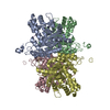
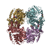
| Entry | Database: PDB / ID: 1ado | ||||||
|---|---|---|---|---|---|---|---|
| Title | FRUCTOSE 1,6-BISPHOSPHATE ALDOLASE FROM RABBIT MUSCLE | ||||||
 Components Components | ALDOLASE Fructose-bisphosphate aldolase Fructose-bisphosphate aldolase | ||||||
 Keywords Keywords | LYASE / ALDOLASE / LYASE (ALDEHYDE) / SCHIFF BASE / GLYCOLYSIS | ||||||
| Function / homology |  Function and homology information Function and homology informationnegative regulation of Arp2/3 complex-mediated actin nucleation / fructose-bisphosphate aldolase / fructose-bisphosphate aldolase activity / M band / I band / glycolytic process / protein homotetramerization / positive regulation of cell migrationSimilarity search - Function | ||||||
| Biological species |  Oryctolagus cuniculus (rabbit) Oryctolagus cuniculus (rabbit) | ||||||
| Method | X-RAY DIFFRACTION / MOLECULAR REPLACEMENT / Resolution: 1.9 Å | ||||||
 Authors Authors | Blom, N.S. / Sygusch, J. | ||||||
 Citation Citation | Journal: Nat.Struct.Biol. / Year: 1997 Title: Product binding and role of the C-terminal region in class I D-fructose 1,6-bisphosphate aldolase. Authors: Blom, N. / Sygusch, J. #1: Journal: To be PublishedTitle: Enhanced Electron Density Envelopes by Extended Solvent Definition Authors: Blom, N.S. / Sygusch, J. | ||||||
| History |
|
- Structure visualization
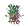
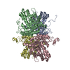
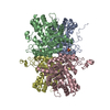
| Structure viewer | Molecule: MolmilJmol/JSmol |
|---|
- Downloads & links
-Download
| PDBx/mmCIF format | 1ado.cif.gz | 417.8 KB | Display | PDBx/mmCIF format |
|---|---|---|---|---|
| PDB format | pdb1ado.ent.gz | 335.6 KB | Display | PDB format |
| PDBx/mmJSON format | 1ado.json.gz | Tree view | PDBx/mmJSON format | |
| Others |  Other downloads Other downloads |
-Validation report
| Arichive directory | https://data.pdbj.org/pub/pdb/validation_reports/ad/1adoftp://data.pdbj.org/pub/pdb/validation_reports/ad/1ado | HTTPS FTP |
|---|
-Related structure data
| Similar structure data |
|---|

-Links
 PDBj
PDBj
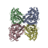
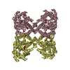
- Assembly
| Deposited unit | 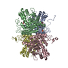
| ||||||||
|---|---|---|---|---|---|---|---|---|---|
| 1 |
| ||||||||
| Unit cell |
|
-Components
| #1: Protein | Fructose-bisphosphate aldolase Mass: 39253.637 Da / Num. of mol.: 4 / Source method: isolated from a natural source / Source: (natural) Oryctolagus cuniculus (rabbit) / Tissue: MUSCLESkeletal muscle / References: UniProt: P00883, fructose-bisphosphate aldolase#2: Chemical | Dihydroxyacetone phosphate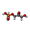  Mass: 170.058 Da / Num. of mol.: 2 / Source method: obtained synthetically / Formula: C3H7O6P Mass: 170.058 Da / Num. of mol.: 2 / Source method: obtained synthetically / Formula: C3H7O6P#3: Chemical | Sulfate  Mass: 96.063 Da / Num. of mol.: 2 / Source method: obtained synthetically / Formula: SO4 Mass: 96.063 Da / Num. of mol.: 2 / Source method: obtained synthetically / Formula: SO4#4: Water | ChemComp-HOH / | Water Mass: 18.015 Da / Num. of mol.: 3288 / Source method: isolated from a natural source / Formula: H2O Mass: 18.015 Da / Num. of mol.: 3288 / Source method: isolated from a natural source / Formula: H2O |
|---|
-Experimental details
-Experiment
| Experiment | Method: X-RAY DIFFRACTION / Number of used crystals: 1 |
|---|
- Sample preparation
| Crystal | Density Matthews: 2.49 Å3/Da / Density % sol: 50.54 % | |||||||||||||||||||||||||
|---|---|---|---|---|---|---|---|---|---|---|---|---|---|---|---|---|---|---|---|---|---|---|---|---|---|---|
| Crystal grow | pH: 7.5 Details: RABBIT MUSCLE ALDOLASE WAS CRYSTALLIZED FROM A 42% SATURATED AMMONIUM SULFATE SOLUTION, pH 7.5 | |||||||||||||||||||||||||
| Crystal grow | *PLUS pH: 7.3 / Method: unknown / Details: Eagles, P. A., (1969) J. Mol. Biol., 45, 533-544 | |||||||||||||||||||||||||
| Components of the solutions | *PLUS
|
-Data collection
| Diffraction | Mean temperature: 298 K |
|---|---|
| Diffraction source | Wavelength: 1.5418 |
| Detector | Type: SIEMENS / Detector: AREA DETECTOR / Date: Oct 1, 1991 |
| Radiation | Monochromator: GRAPHITE(002) / Monochromatic (M) / Laue (L): M / Scattering type: x-ray |
| Radiation wavelength | Wavelength: 1.5418 Å / Relative weight: 1 |
| Reflection | Resolution: 1.9→12 Å / Num. obs: 103662 / % possible obs: 85.2 % / Observed criterion σ(I): 1 / Rsym value: 0.056 / Net I/σ(I): 14.5 |
| Reflection shell | Resolution: 1.9→2 Å / Mean I/σ(I) obs: 1.9 / Rsym value: 0.286 |
| Reflection | *PLUS Rmerge(I) obs: 0.056 |
| Reflection shell | *PLUS Rmerge(I) obs: 0.286 |
- Processing
| Software |
| ||||||||||||||||||||||||||||||||||||||||||||||||||||||||||||
|---|---|---|---|---|---|---|---|---|---|---|---|---|---|---|---|---|---|---|---|---|---|---|---|---|---|---|---|---|---|---|---|---|---|---|---|---|---|---|---|---|---|---|---|---|---|---|---|---|---|---|---|---|---|---|---|---|---|---|---|---|---|
| Refinement | Method to determine structure: MOLECULAR REPLACEMENT Starting model: STRUCTURE TO 2.7 ANGSTROM RESOLUTION Resolution: 1.9→12 Å / Data cutoff high absF: 100000 / Data cutoff low absF: 0.1 / Isotropic thermal model: RESTRAINED / Cross valid method: THROUGHOUT / σ(F): 1
| ||||||||||||||||||||||||||||||||||||||||||||||||||||||||||||
| Displacement parameters | Biso mean: 29.1 Å2
| ||||||||||||||||||||||||||||||||||||||||||||||||||||||||||||
| Refinement step | Cycle: LAST / Resolution: 1.9→12 Å
| ||||||||||||||||||||||||||||||||||||||||||||||||||||||||||||
| Refine LS restraints |
| ||||||||||||||||||||||||||||||||||||||||||||||||||||||||||||
| LS refinement shell | Resolution: 1.9→1.99 Å / Total num. of bins used: 8
| ||||||||||||||||||||||||||||||||||||||||||||||||||||||||||||
| Xplor file |
| ||||||||||||||||||||||||||||||||||||||||||||||||||||||||||||
| Software | *PLUS Name: X-PLOR / Version: 3.1 / Classification: refinement | ||||||||||||||||||||||||||||||||||||||||||||||||||||||||||||
| Refinement | *PLUS | ||||||||||||||||||||||||||||||||||||||||||||||||||||||||||||
| Solvent computation | *PLUS | ||||||||||||||||||||||||||||||||||||||||||||||||||||||||||||
| Displacement parameters | *PLUS | ||||||||||||||||||||||||||||||||||||||||||||||||||||||||||||
| Refine LS restraints | *PLUS
| ||||||||||||||||||||||||||||||||||||||||||||||||||||||||||||
| LS refinement shell | *PLUS Rfactor obs: 0.262 |
