 Movie
Movie Controller


+ Open data

- Basic information
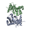
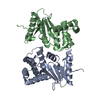
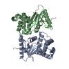
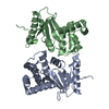
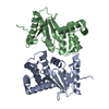
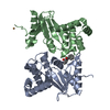
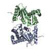
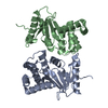
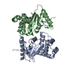
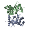
| Entry | Database: PDB / ID: 1ac1 | ||||||
|---|---|---|---|---|---|---|---|
| Title | DSBA MUTANT H32L | ||||||
 Components Components | DSBA | ||||||
 Keywords Keywords | DISULFIDE OXIDOREDUCTASE / THIOREDOXIN FOLD / REDOX-ACTIVE CENTER | ||||||
| Function / homology |  Function and homology information Function and homology informationcellular response to antibiotic / protein disulfide isomerase activity / protein-disulfide reductase activity / outer membrane-bounded periplasmic space / periplasmic space / oxidoreductase activitySimilarity search - Function | ||||||
| Biological species |  Escherichia coli (E. coli) Escherichia coli (E. coli) | ||||||
| Method | X-RAY DIFFRACTION / DIFFERENCE FOURIER / Resolution: 2 Å | ||||||
 Authors Authors | Guddat, L.W. / Martin, J.L. | ||||||
 Citation Citation | Journal: Protein Sci. / Year: 1997 Title: Structural analysis of three His32 mutants of DsbA: support for an electrostatic role of His32 in DsbA stability. Authors: Guddat, L.W. / Bardwell, J.C. / Glockshuber, R. / Huber-Wunderlich, M. / Zander, T. / Martin, J.L. #1: Journal: Protein Sci. / Year: 1997Title: The Uncharged Surface Features Surrounding the Active Site of Escherichia Coli Dsba are Conserved and are Implicated in Peptide Binding Authors: Guddat, L.W. / Bardwell, J.C. / Zander, T. / Martin, J.L. #2: Journal: Nature / Year: 1993Title: Crystal Structure of the Dsba Protein Required for Disulphide Bond Formation in Vivo Authors: Martin, J.L. / Bardwell, J.C. / Kuriyan, J. | ||||||
| History |
|
- Structure visualization
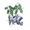
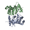
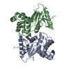
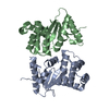
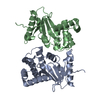
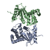
| Structure viewer | Molecule: MolmilJmol/JSmol |
|---|
- Downloads & links
-Download
| PDBx/mmCIF format | 1ac1.cif.gz | 82.1 KB | Display | PDBx/mmCIF format |
|---|---|---|---|---|
| PDB format | pdb1ac1.ent.gz | 65.5 KB | Display | PDB format |
| PDBx/mmJSON format | 1ac1.json.gz | Tree view | PDBx/mmJSON format | |
| Others |  Other downloads Other downloads |
-Validation report
| Arichive directory | https://data.pdbj.org/pub/pdb/validation_reports/ac/1ac1ftp://data.pdbj.org/pub/pdb/validation_reports/ac/1ac1 | HTTPS FTP |
|---|
-Related structure data
| Related structure data | 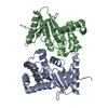 1acvC 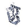 1fvjC  1fvkSC S: Starting model for refinement C: citing same article ( |
|---|---|
| Similar structure data |


-Links
 PDBj
PDBj

- Assembly
| Deposited unit | 
| ||||||||
|---|---|---|---|---|---|---|---|---|---|
| 1 |
| ||||||||
| Unit cell |
| ||||||||
| Noncrystallographic symmetry (NCS) | NCS oper: (Code: given Matrix: (-0.962443, 0.133572, 0.236352), Vector : |
-Components
| #1: Protein | / DISULFIDE BOND FORMATION PROTEIN Mass: 21130.035 Da / Num. of mol.: 2 / Mutation: H32L Source method: isolated from a genetically manipulated source Source: (gene. exp.) Escherichia coli (E. coli) / Cellular location: PERIPLASM / Production host: Escherichia coli (E. coli) / References: UniProt: P24991, UniProt: P0AEG4*PLUS#2: Water | ChemComp-HOH / | Water Mass: 18.015 Da / Num. of mol.: 157 / Source method: isolated from a natural source / Formula: H2O Mass: 18.015 Da / Num. of mol.: 157 / Source method: isolated from a natural source / Formula: H2O |
|---|
-Experimental details
-Experiment
| Experiment | Method: X-RAY DIFFRACTION / Number of used crystals: 1 |
|---|
- Sample preparation
| Crystal | Density Matthews: 2.8 Å3/Da / Density % sol: 53.1 % | ||||||||||||||||||||||||||||||||||||||||||||||||
|---|---|---|---|---|---|---|---|---|---|---|---|---|---|---|---|---|---|---|---|---|---|---|---|---|---|---|---|---|---|---|---|---|---|---|---|---|---|---|---|---|---|---|---|---|---|---|---|---|---|
| Crystal grow | pH: 6.3 / Details: CACODYLATE PH 6.3 PEG 8K 25% | ||||||||||||||||||||||||||||||||||||||||||||||||
| Crystal grow | *PLUS Temperature: 21 ℃ / pH: 6.5 / Method: vapor diffusion, hanging drop / Details: Martin, J.L., (1993) J.Mol.Biol., 230, 1097. | ||||||||||||||||||||||||||||||||||||||||||||||||
| Components of the solutions | *PLUS
|
-Data collection
| Diffraction | Mean temperature: 290 K |
|---|---|
| Diffraction source | Source: ROTATING ANODE / Type: RIGAKU RUH2R / Wavelength: 1.5418 |
| Detector | Type: RIGAKU RAXIS IIC / Detector: IMAGE PLATE / Date: Sep 1, 1996 / Details: MIRRORS |
| Radiation | Monochromator: NI FILTER / Monochromatic (M) / Laue (L): M / Scattering type: x-ray |
| Radiation wavelength | Wavelength: 1.5418 Å / Relative weight: 1 |
| Reflection | Resolution: 2→50 Å / Num. obs: 28490 / % possible obs: 91.5 % / Observed criterion σ(I): 1 / Redundancy: 2.9 % / Biso Wilson estimate: 27.2 Å2 / Rmerge(I) obs: 0.081 / Net I/σ(I): 13.6 |
| Reflection shell | Resolution: 2→2.07 Å / Redundancy: 2.83 % / Rmerge(I) obs: 0.298 / Mean I/σ(I) obs: 2.36 / % possible all: 83 |
| Reflection | *PLUS Num. measured all: 84044 |
| Reflection shell | *PLUS Highest resolution: 2 Å / % possible obs: 83 % |
- Processing
| Software |
| ||||||||||||||||||||||||||||||||||||||||||||||||||||||||||||
|---|---|---|---|---|---|---|---|---|---|---|---|---|---|---|---|---|---|---|---|---|---|---|---|---|---|---|---|---|---|---|---|---|---|---|---|---|---|---|---|---|---|---|---|---|---|---|---|---|---|---|---|---|---|---|---|---|---|---|---|---|---|
| Refinement | Method to determine structure: DIFFERENCE FOURIER Starting model: 1.7 A STRUCTURE OF WILDTYPE OXIDISED DSBA (PDB ENTRY 1FVK) Resolution: 2→50 Å / Data cutoff low absF: 0.1 / Isotropic thermal model: RESTRAINED / Cross valid method: THROUGHOUT / σ(F): 1 Details: BULK SOLVENT CORRECTION USED SOLDEN=0.34, SOLRAD = 0.25 A
| ||||||||||||||||||||||||||||||||||||||||||||||||||||||||||||
| Displacement parameters | Biso mean: 33.5 Å2 | ||||||||||||||||||||||||||||||||||||||||||||||||||||||||||||
| Refine analyze |
| ||||||||||||||||||||||||||||||||||||||||||||||||||||||||||||
| Refinement step | Cycle: LAST / Resolution: 2→50 Å
| ||||||||||||||||||||||||||||||||||||||||||||||||||||||||||||
| Refine LS restraints |
| ||||||||||||||||||||||||||||||||||||||||||||||||||||||||||||
| LS refinement shell | Resolution: 2→2.09 Å / Total num. of bins used: 8
| ||||||||||||||||||||||||||||||||||||||||||||||||||||||||||||
| Xplor file |
| ||||||||||||||||||||||||||||||||||||||||||||||||||||||||||||
| Software | *PLUS Name: X-PLOR / Version: 3.1 / Classification: refinement | ||||||||||||||||||||||||||||||||||||||||||||||||||||||||||||
| Refinement | *PLUS Rfactor Rfree: 0.22 | ||||||||||||||||||||||||||||||||||||||||||||||||||||||||||||
| Solvent computation | *PLUS | ||||||||||||||||||||||||||||||||||||||||||||||||||||||||||||
| Displacement parameters | *PLUS | ||||||||||||||||||||||||||||||||||||||||||||||||||||||||||||
| Refine LS restraints | *PLUS
|
