 Movie
Movie Controller


+ Open data

- Basic information
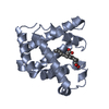
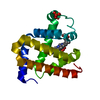
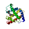
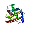
| Entry | Database: PDB / ID: 1abs | ||||||
|---|---|---|---|---|---|---|---|
| Title | PHOTOLYSED CARBONMONOXY-MYOGLOBIN AT 20 K | ||||||
 Components Components | MYOGLOBIN | ||||||
 Keywords Keywords | OXYGEN STORAGE / INTERMEDIATE IN LIGAND BINDING / RESPIRATORY PROTEIN | ||||||
| Function / homology |  Function and homology information Function and homology informationhydrogen peroxide mediated signaling pathway / oxygen carrier activity / oxygen binding / heme binding / metal ion bindingSimilarity search - Function | ||||||
| Biological species |  Physeter catodon (sperm whale) Physeter catodon (sperm whale) | ||||||
| Method | X-RAY DIFFRACTION / SYNCHROTRON / DIFFERENCE FOURIER / Resolution: 1.5 Å | ||||||
 Authors Authors | Schlichting, I. / Berendzen, J. / Phillips Jr., G.N. / Sweet, R.M. | ||||||
 Citation Citation | Journal: Nature / Year: 1994 Title: Crystal structure of photolysed carbonmonoxy-myoglobin. Authors: Schlichting, I. / Berendzen, J. / Phillips Jr., G.N. / Sweet, R.M. #1: Journal: Proteins / Year: 1990Title: Crystal Structure of Myoglobin from a Synthetic Gene Authors: Phillips Jr., G.N. / Arduini, R.M. / Springer, B.A. / Sligar, S.G. #2: Journal: Proc.Natl.Acad.Sci.USA / Year: 1987Title: High-Level Expression of Sperm Whale Myoglobin in Escherichia Coli Authors: Springer, B.A. / Sligar, S.G. | ||||||
| History |
|
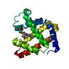
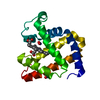
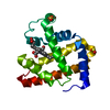
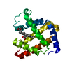
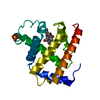
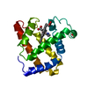
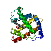
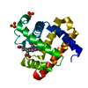
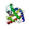
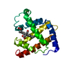
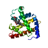
- Structure visualization
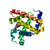
| Structure viewer | Molecule: MolmilJmol/JSmol |
|---|
- Downloads & links
-Download
| PDBx/mmCIF format | 1abs.cif.gz | 43.6 KB | Display | PDBx/mmCIF format |
|---|---|---|---|---|
| PDB format | pdb1abs.ent.gz | 33.2 KB | Display | PDB format |
| PDBx/mmJSON format | 1abs.json.gz | Tree view | PDBx/mmJSON format | |
| Others |  Other downloads Other downloads |
-Validation report
| Arichive directory | https://data.pdbj.org/pub/pdb/validation_reports/ab/1absftp://data.pdbj.org/pub/pdb/validation_reports/ab/1abs | HTTPS FTP |
|---|
-Related structure data
| Related structure data | 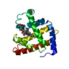 2mgkS S: Starting model for refinement |
|---|---|
| Similar structure data |


-Links
 PDBj
PDBj


- Assembly
| Deposited unit | 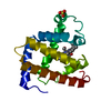
| ||||||||
|---|---|---|---|---|---|---|---|---|---|
| 1 |
| ||||||||
| Unit cell |
|
-Components
| #1: Protein | Mass: 17365.164 Da / Num. of mol.: 1 / Mutation: D122N Source method: isolated from a genetically manipulated source Details: HEME BOUND TO HIS-93, PHOTOLYSED CO ATOP HEME / Source: (gene. exp.) Physeter catodon (sperm whale) / Description: SYNTHETIC GENE / Production host:  Escherichia coli (E. coli) / References: UniProt: P02185 Escherichia coli (E. coli) / References: UniProt: P02185 |
|---|---|
| #2: Chemical | ChemComp-SO4 / Sulfate  Mass: 96.063 Da / Num. of mol.: 1 / Source method: obtained synthetically / Formula: SO4 Mass: 96.063 Da / Num. of mol.: 1 / Source method: obtained synthetically / Formula: SO4 |
| #3: Chemical | ChemComp-HEM / Heme B  Mass: 616.487 Da / Num. of mol.: 1 / Source method: obtained synthetically / Formula: C34H32FeN4O4 Mass: 616.487 Da / Num. of mol.: 1 / Source method: obtained synthetically / Formula: C34H32FeN4O4 |
| #4: Chemical | ChemComp-CMO / Carbon monoxide  Mass: 28.010 Da / Num. of mol.: 1 / Source method: obtained synthetically / Formula: CO Mass: 28.010 Da / Num. of mol.: 1 / Source method: obtained synthetically / Formula: CO |
| #5: Water | ChemComp-HOH / Water Mass: 18.015 Da / Num. of mol.: 118 / Source method: isolated from a natural source / Formula: H2O Mass: 18.015 Da / Num. of mol.: 118 / Source method: isolated from a natural source / Formula: H2O |
-Experimental details
-Experiment
| Experiment | Method: X-RAY DIFFRACTION / Number of used crystals: 2 |
|---|
- Sample preparation
| Crystal | Density Matthews: 3.08 Å3/Da / Density % sol: 60.12 % Description: DATA WERE COLLECTED UNDER CONTINUOUS ILLUMINATION USING AN OPEN FLOW HELIUM CRYOSTAT FOR COOLING. | |||||||||||||||||||||||||
|---|---|---|---|---|---|---|---|---|---|---|---|---|---|---|---|---|---|---|---|---|---|---|---|---|---|---|
| Crystal grow | pH: 9 Details: PROTEIN WAS CRYSTALLIZED FROM 3.6 M AMMONIUM SULFATE. CRYSTALS WER SOAKED FOR 1 HOUR IN A THOROUGHLY DEGASSED AND N2 - SATURATED CRYOPROTECTANT SOLUTION MADE BY ADDITION OF 100 MG SUCROSE, ...Details: PROTEIN WAS CRYSTALLIZED FROM 3.6 M AMMONIUM SULFATE. CRYSTALS WER SOAKED FOR 1 HOUR IN A THOROUGHLY DEGASSED AND N2 - SATURATED CRYOPROTECTANT SOLUTION MADE BY ADDITION OF 100 MG SUCROSE, 100 MG GLUCOSE AND 8 MG NA-DITHIONATE TO 1 ML OF 70% SATURATED AMMONIUM SULFATE WITH 50 MM TRIS. HCL PH 9.0. THEN THE SOLUTION WAS EXCHANGED AGAINST A CO SATURATED ONE. DISTINCT COLOR CHANGES ACCOMPANIED THE FORMATION OF UNLIGATED MYOGLOBIN FROM MET-MB AND MBCO FROM UNLIGATED MB. | |||||||||||||||||||||||||
| Crystal grow | *PLUS Method: batch method | |||||||||||||||||||||||||
| Components of the solutions | *PLUS
|
-Data collection
| Diffraction | Mean temperature: 20 K |
|---|---|
| Diffraction source | Source: SYNCHROTRON / Site: NSLS  / Beamline: X12C / Wavelength: 0.91 / Beamline: X12C / Wavelength: 0.91 |
| Detector | Type: MARRESEARCH / Detector: IMAGE PLATE AREA DETECTOR / Date: Mar 1, 1994 / Details: YES |
| Radiation | Monochromator: YES / Monochromatic (M) / Laue (L): M / Scattering type: x-ray |
| Radiation wavelength | Wavelength: 0.91 Å / Relative weight: 1 |
| Reflection | Highest resolution: 1.5 Å / Num. obs: 30947 / % possible obs: 90 % / Observed criterion σ(I): 0 / Redundancy: 3 % / Rmerge(I) obs: 0.063 / Rsym value: 0.068 |
| Reflection shell | Resolution: 1.5→1.7 Å / % possible all: 78 |
| Reflection | *PLUS Lowest resolution: 9999 Å / Num. measured all: 93946 / Rmerge(I) obs: 0.063 |
| Reflection shell | *PLUS % possible obs: 78 % |
- Processing
| Software |
| ||||||||||||||||||||||||||||||||||||||||||||||||||||||||||||
|---|---|---|---|---|---|---|---|---|---|---|---|---|---|---|---|---|---|---|---|---|---|---|---|---|---|---|---|---|---|---|---|---|---|---|---|---|---|---|---|---|---|---|---|---|---|---|---|---|---|---|---|---|---|---|---|---|---|---|---|---|---|
| Refinement | Method to determine structure: DIFFERENCE FOURIER Starting model: 2MGK Resolution: 1.5→7 Å / Data cutoff high absF: 10000000 / Data cutoff low absF: 0 / σ(F): 0 Details: HEME PARAMETERS WERE RELAXED TO ALLOW FOR REFINEMENT OF UNLIGATED, PHOTOLYSED, AND CO-BOUND COMPLEX.
| ||||||||||||||||||||||||||||||||||||||||||||||||||||||||||||
| Displacement parameters | Biso mean: 6.4 Å2 | ||||||||||||||||||||||||||||||||||||||||||||||||||||||||||||
| Refine analyze | Luzzati d res low obs: 7 Å / Luzzati sigma a obs: 0.06 Å | ||||||||||||||||||||||||||||||||||||||||||||||||||||||||||||
| Refinement step | Cycle: LAST / Resolution: 1.5→7 Å
| ||||||||||||||||||||||||||||||||||||||||||||||||||||||||||||
| Refine LS restraints |
| ||||||||||||||||||||||||||||||||||||||||||||||||||||||||||||
| Refine LS restraints NCS | NCS model details: N.A | ||||||||||||||||||||||||||||||||||||||||||||||||||||||||||||
| LS refinement shell | Resolution: 1.5→1.57 Å / Total num. of bins used: 8
| ||||||||||||||||||||||||||||||||||||||||||||||||||||||||||||
| Xplor file |
| ||||||||||||||||||||||||||||||||||||||||||||||||||||||||||||
| Software | *PLUS Name: X-PLOR / Classification: refinement | ||||||||||||||||||||||||||||||||||||||||||||||||||||||||||||
| Refinement | *PLUS | ||||||||||||||||||||||||||||||||||||||||||||||||||||||||||||
| Solvent computation | *PLUS | ||||||||||||||||||||||||||||||||||||||||||||||||||||||||||||
| Displacement parameters | *PLUS | ||||||||||||||||||||||||||||||||||||||||||||||||||||||||||||
| LS refinement shell | *PLUS Rfactor all: 0.263 |
