 Movie
Movie Controller


+ Open data

- Basic information
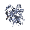
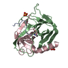
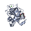
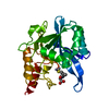
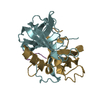
| Entry | Database: PDB / ID: 1ab9 | ||||||
|---|---|---|---|---|---|---|---|
| Title | CRYSTAL STRUCTURE OF BOVINE GAMMA-CHYMOTRYPSIN | ||||||
 Components Components |
| ||||||
 Keywords Keywords | COMPLEX (SERINE PROTEASE/PEPTIDE) /  HYDROLASE / SERINE PROTEASE / DIGESTION / PANCREAS / ZYMOGEN / COMPLEX (SERINE PROTEASE-PEPTIDE) / COMPLEX (SERINE PROTEASE-PEPTIDE) complex HYDROLASE / SERINE PROTEASE / DIGESTION / PANCREAS / ZYMOGEN / COMPLEX (SERINE PROTEASE-PEPTIDE) / COMPLEX (SERINE PROTEASE-PEPTIDE) complex | ||||||
| Function / homology |  Function and homology informationchymotrypsin / serpin family protein binding / serine protease inhibitor complex / digestion / serine-type endopeptidase activity / proteolysis / extracellular region Function and homology informationchymotrypsin / serpin family protein binding / serine protease inhibitor complex / digestion / serine-type endopeptidase activity / proteolysis / extracellular regionSimilarity search - Function | ||||||
| Biological species |  Bos taurus (cattle) Bos taurus (cattle) | ||||||
| Method | X-RAY DIFFRACTION / MOLECULAR REPLACEMENT / Resolution: 1.6 Å | ||||||
 Authors Authors | Sugio, S. / Kashima, A. / Inoue, Y. / Maeda, I. / Nose, T. / Shimohigashi, Y. | ||||||
 Citation Citation | Journal: Eur.J.Biochem. / Year: 1998 Title: X-ray crystal structure of a dipeptide-chymotrypsin complex in an inhibitory interaction. Authors: Kashima, A. / Inoue, Y. / Sugio, S. / Maeda, I. / Nose, T. / Shimohigashi, Y. #1: Journal: Biochemistry / Year: 1991Title: Gamma-Chymotrypsin is a Complex of Alpha-Chymotrypsin with its Own Autolysis Products Authors: Harel, M. / Su, C.T. / Frolow, F. / Silman, I. / Sussman, J.L. #2: Journal: Int.J.Biol.Macromol. / Year: 1991Title: Structure of Gamma-Chymotrypsin in the Range Ph 2.0 To Ph 10.5 Suggests that Gamma-Chymotrypsin is a Covalent Acyl-Enzyme Adduct at Low Ph Authors: Dixon, M.M. / Brennan, R.G. / Matthews, B.W. #3: Journal: Biochemistry / Year: 1989Title: Is Gamma-Chymotrypsin a Tetrapeptide Acyl-Enzyme Adduct of Alpha-Chymotrypsin? Authors: Dixon, M.M. / Matthews, B.W. | ||||||
| History |
|
- Structure visualization
| Structure viewer | Molecule: MolmilJmol/JSmol |
|---|
- Downloads & links
-Download
| PDBx/mmCIF format | 1ab9.cif.gz | 58.4 KB | Display | PDBx/mmCIF format |
|---|---|---|---|---|
| PDB format | pdb1ab9.ent.gz | 44.9 KB | Display | PDB format |
| PDBx/mmJSON format | 1ab9.json.gz | Tree view | PDBx/mmJSON format | |
| Others |  Other downloads Other downloads |
-Validation report
| Arichive directory | https://data.pdbj.org/pub/pdb/validation_reports/ab/1ab9ftp://data.pdbj.org/pub/pdb/validation_reports/ab/1ab9 | HTTPS FTP |
|---|
-Related structure data
| Related structure data |  1afqC  1gctS S: Starting model for refinement C: citing same article ( |
|---|---|
| Similar structure data |


-Links
 PDBj
PDBj

- Assembly
| Deposited unit | 
| |||||||||
|---|---|---|---|---|---|---|---|---|---|---|
| 1 |
| |||||||||
| 2 | 
| |||||||||
| 3 | 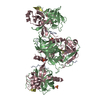
| |||||||||
| Unit cell |
| |||||||||
| Components on special symmetry positions |
|
-Components
-Protein/peptide , 2 types, 2 molecules AD
| #1: Protein/peptide | Mass: 1253.511 Da / Num. of mol.: 1 / Source method: isolated from a natural source / Source: (natural) Bos taurus (cattle) / References: UniProt: P00766, chymotrypsin |
|---|---|
| #4: Protein/peptide | Mass: 535.590 Da / Num. of mol.: 1 Source method: isolated from a genetically manipulated source |
-Protein , 2 types, 2 molecules BC
| #2: Protein | Mass: 13934.556 Da / Num. of mol.: 1 / Source method: isolated from a natural source / Source: (natural) Bos taurus (cattle) / References: UniProt: P00766, chymotrypsin |
|---|---|
| #3: Protein | Mass: 10074.495 Da / Num. of mol.: 1 / Source method: isolated from a natural source / Source: (natural) Bos taurus (cattle) / References: UniProt: P00766, chymotrypsin |
-Non-polymers , 2 types, 128 molecules 

| #5: Chemical | ChemComp-SO4 / Sulfate Mass: 96.063 Da / Num. of mol.: 1 / Source method: obtained synthetically / Formula: SO4 Mass: 96.063 Da / Num. of mol.: 1 / Source method: obtained synthetically / Formula: SO4 |
|---|---|
| #6: Water | ChemComp-HOH / WaterMass: 18.015 Da / Num. of mol.: 127 / Source method: isolated from a natural source / Formula: H2O |
-Details
| Compound details | THE BOUND PENTAPEPTIDE (THR D 300 TO TYR D 304) HAVE TWO DIFFERENT CONFORMATIONS. THE OCCUPANCIES ...THE BOUND PENTAPEPTI |
|---|
-Experimental details
-Experiment
| Experiment | Method: X-RAY DIFFRACTION / Number of used crystals: 1 |
|---|
- Sample preparation
| Crystal | Density Matthews: 2.29 Å3/Da / Density % sol: 46.28 % | ||||||||||||||||||||||||||||||
|---|---|---|---|---|---|---|---|---|---|---|---|---|---|---|---|---|---|---|---|---|---|---|---|---|---|---|---|---|---|---|---|
| Crystal grow | Method: vapor diffusion, sitting drop / pH: 5.6 Details: ALPHA-CHYMOTRYPSIN WAS DISSOLVED IN POTASSIUM BORATE (PH8.6) AND WAS INCUBATED AT 310K FOR 6 HOURS. SOLID AMMONIUM SULFATE WAS ADDED TO THE SOLUTION, AND THE PRECIPITATE FORMED WAS RECOVERED ...Details: ALPHA-CHYMOTRYPSIN WAS DISSOLVED IN POTASSIUM BORATE (PH8.6) AND WAS INCUBATED AT 310K FOR 6 HOURS. SOLID AMMONIUM SULFATE WAS ADDED TO THE SOLUTION, AND THE PRECIPITATE FORMED WAS RECOVERED AND RE-DISSOLVED WITH WATER. CRYSTALLIZATION WAS DONE WITH A SITTING-DROP VAPOR-DIFFUSION PROCEDURE, IN WHICH PROTEIN SOLUTION (15MG/ML) CONTAINING 10MM CACODYLATE, 0.75% SATURATED ACETYLTRIMETHYL AMMONIUM AND 45% SATURATED AMMONIUM SULFATE WAS EQUILIBRATED AGAINST 65% SATURATED AMMONIUM SULFATE AT 293K., pH 5.6, vapor diffusion - sitting drop | ||||||||||||||||||||||||||||||
| Crystal grow | *PLUS Method: vapor diffusion | ||||||||||||||||||||||||||||||
| Components of the solutions | *PLUS
|
-Data collection
| Diffraction | Mean temperature: 292 K |
|---|---|
| Diffraction source | Source: ROTATING ANODE / Type: RIGAKU RUH2R / Wavelength: 1.5418 |
| Detector | Type: RIGAKU RAXIS IIC / Detector: IMAGE PLATE / Date: Oct 24, 1995 / Details: YALE MIRRORS |
| Radiation | Monochromator: NI FILTER / Monochromatic (M) / Laue (L): M / Scattering type: x-ray |
| Radiation wavelength | Wavelength: 1.5418 Å / Relative weight: 1 |
| Reflection | Resolution: 1.6→69.5 Å / Num. obs: 31740 / % possible obs: 97.8 % / Observed criterion σ(I): 1 / Redundancy: 10.3 % / Biso Wilson estimate: 19 Å2 / Rmerge(I) obs: 0.055 / Net I/σ(I): 15.1 |
| Reflection shell | Resolution: 1.6→1.65 Å / Rmerge(I) obs: 0.208 / Mean I/σ(I) obs: 3.1 / % possible all: 94.7 |
| Reflection | *PLUS Num. measured all: 332021 |
| Reflection shell | *PLUS % possible obs: 94.7 % |
- Processing
| Software |
| ||||||||||||||||||||||||||||||||||||||||||||||||||||||||||||
|---|---|---|---|---|---|---|---|---|---|---|---|---|---|---|---|---|---|---|---|---|---|---|---|---|---|---|---|---|---|---|---|---|---|---|---|---|---|---|---|---|---|---|---|---|---|---|---|---|---|---|---|---|---|---|---|---|---|---|---|---|---|
| Refinement | Method to determine structure: MOLECULAR REPLACEMENT Starting model: PDB ENTRY 1GCT Resolution: 1.6→5 Å / Rfactor Rfree error: 0.004 / Data cutoff high absF: 100000 / Data cutoff low absF: 0.1 / Isotropic thermal model: RESTRAINED / Cross valid method: POSTERIORI / σ(F): 2 Details: THE FOLLOWING WEIGHTING SCHEME WAS USED: 1/(SIGMAF)**2 SIDE CHAINS OF VAL B 53 AND GLN C 239 HAVE ALTERNATE CONFORMATIONS. THE OCCUPANCIES OF THE CORRESPONDING ATOMS WERE SET TO 0.5.
| ||||||||||||||||||||||||||||||||||||||||||||||||||||||||||||
| Displacement parameters | Biso mean: 21.2 Å2 | ||||||||||||||||||||||||||||||||||||||||||||||||||||||||||||
| Refine analyze | Luzzati coordinate error obs: 0.2 Å / Luzzati d res low obs: 5 Å | ||||||||||||||||||||||||||||||||||||||||||||||||||||||||||||
| Refinement step | Cycle: LAST / Resolution: 1.6→5 Å
| ||||||||||||||||||||||||||||||||||||||||||||||||||||||||||||
| Refine LS restraints |
| ||||||||||||||||||||||||||||||||||||||||||||||||||||||||||||
| LS refinement shell | Resolution: 1.6→1.66 Å / Rfactor Rfree error: 0.017 / Total num. of bins used: 10
| ||||||||||||||||||||||||||||||||||||||||||||||||||||||||||||
| Xplor file |
| ||||||||||||||||||||||||||||||||||||||||||||||||||||||||||||
| Software | *PLUS Name: X-PLOR / Version: 3.1 / Classification: refinement | ||||||||||||||||||||||||||||||||||||||||||||||||||||||||||||
| Refinement | *PLUS | ||||||||||||||||||||||||||||||||||||||||||||||||||||||||||||
| Solvent computation | *PLUS | ||||||||||||||||||||||||||||||||||||||||||||||||||||||||||||
| Displacement parameters | *PLUS | ||||||||||||||||||||||||||||||||||||||||||||||||||||||||||||
| Refine LS restraints | *PLUS
| ||||||||||||||||||||||||||||||||||||||||||||||||||||||||||||
| LS refinement shell | *PLUS Rfactor obs: 0.285 |
