Journal: Am.Cryst.Assoc.,Abstr.Papers (Annual Meeting) / Year: 1998 Title: The 1.61 Angstrom Structure of the Tetanus Toxin Ganglioside Binding Region: Solved by MAD and Mir Phase Combination Authors: Knapp, M. / Segelke, B. / Rupp, B.
Mass: 18.015 Da / Num. of mol.: 424 / Source method: isolated from a natural source / Formula: H2O
-
Experimental details
-
Experiment
Experiment
Method: X-RAY DIFFRACTION / Number of used crystals: 1
-
Sample preparation
Crystal
Density Matthews: 2.21 Å3/Da / Density % sol: 44.4 %
Crystal grow
Method: fiber seeding using cryshed initial crystals as seeds pH: 7 Details: 9% (W/V) 1M AMMONIUM PHOSPHATE, PH 6.0, AND 92% OF A 10% (W/V) SOLUTION OF PEG 6000 IN 50MM HEPES (PH 7.0). LARGE CRYSTALS (0.2 X 0.2 X 0.5 MM) WERE GROWN BY FIBER SEEDING, USING CRUSHED ...Details: 9% (W/V) 1M AMMONIUM PHOSPHATE, PH 6.0, AND 92% OF A 10% (W/V) SOLUTION OF PEG 6000 IN 50MM HEPES (PH 7.0). LARGE CRYSTALS (0.2 X 0.2 X 0.5 MM) WERE GROWN BY FIBER SEEDING, USING CRUSHED INITIAL CRYSTALS AS SEEDS., fiber seeding using cryshed initial crystals as seeds PH range: 6.0-7.0
Highest resolution: 1.57 Å / Num. obs: 72263 / % possible obs: 96.6 % / Redundancy: 4.1 % / Rmerge(I) obs: 0.0569 / Net I/σ(I): 9.6
Reflection shell
Resolution: 1.57→1.6 Å / Redundancy: 2.8 % / Rmerge(I) obs: 0.345 / Mean I/σ(I) obs: 2.05 / % possible all: 90.3
-
Processing
Software
Name
Version
Classification
MOSFLM
datareduction
SCALA
datascaling
SHELXL-97
modelbuilding
SOLVE
phasing
SHELXL-97
refinement
CCP4
(SCALA)
datascaling
SHELXL-97
phasing
Refinement
Method to determine structure: COMBINED MIR (SIRAS), MAD INITIAL REFINEMENT WITH REFMAC, X-PLOR. FINAL REFINEMENT WITH SHELXL. Resolution: 1.57→22.5 Å / Num. parameters: 36750 / Num. restraintsaints: 45026 / Cross valid method: THROUGHOUT / σ(F): 0 / Stereochemistry target values: ENGH AND HUBER Details: ANISOTROPIC REFINEMENT REDUCED FREE R (NO CUTOFF) BY 0.07 (7%)
Rfactor
Num. reflection
% reflection
Selection details
Rfree
0.2618
7306
10.1 %
RANDOM
all
0.1867
72263
-
-
obs
0.1785
-
96.6 %
-
Refine analyze
Num. disordered residues: 0 / Occupancy sum hydrogen: 0 / Occupancy sum non hydrogen: 4082.4
Refinement step
Cycle: LAST / Resolution: 1.57→22.5 Å
Protein
Nucleic acid
Ligand
Solvent
Total
Num. atoms
3657
0
2
424
4083
Refine LS restraints
Refine-ID
Type
Dev ideal
X-RAY DIFFRACTION
s_bond_d
0.008
X-RAY DIFFRACTION
s_angle_d
0.025
X-RAY DIFFRACTION
s_similar_dist
0
X-RAY DIFFRACTION
s_from_restr_planes
0.028
X-RAY DIFFRACTION
s_zero_chiral_vol
0.035
X-RAY DIFFRACTION
s_non_zero_chiral_vol
0.048
X-RAY DIFFRACTION
s_anti_bump_dis_restr
0.014
X-RAY DIFFRACTION
s_rigid_bond_adp_cmpnt
0.007
X-RAY DIFFRACTION
s_similar_adp_cmpnt
0.058
X-RAY DIFFRACTION
s_approx_iso_adps
0.086
+
About Yorodumi
-
News
-
Feb 9, 2022. New format data for meta-information of EMDB entries
New format data for meta-information of EMDB entries
Version 3 of the EMDB header file is now the official format.
The previous official version 1.9 will be removed from the archive.
In the structure databanks used in Yorodumi, some data are registered as the other names, "COVID-19 virus" and "2019-nCoV". Here are the details of the virus and the list of structure data.
Jan 31, 2019. EMDB accession codes are about to change! (news from PDBe EMDB page)
EMDB accession codes are about to change! (news from PDBe EMDB page)
The allocation of 4 digits for EMDB accession codes will soon come to an end. Whilst these codes will remain in use, new EMDB accession codes will include an additional digit and will expand incrementally as the available range of codes is exhausted. The current 4-digit format prefixed with “EMD-” (i.e. EMD-XXXX) will advance to a 5-digit format (i.e. EMD-XXXXX), and so on. It is currently estimated that the 4-digit codes will be depleted around Spring 2019, at which point the 5-digit format will come into force.
The EM Navigator/Yorodumi systems omit the EMD- prefix.
Related info.:Q: What is EMD? / ID/Accession-code notation in Yorodumi/EM Navigator
Yorodumi is a browser for structure data from EMDB, PDB, SASBDB, etc.
This page is also the successor to EM Navigator detail page, and also detail information page/front-end page for Omokage search.
The word "yorodu" (or yorozu) is an old Japanese word meaning "ten thousand". "mi" (miru) is to see.
Related info.:EMDB / PDB / SASBDB / Comparison of 3 databanks / Yorodumi Search / Aug 31, 2016. New EM Navigator & Yorodumi / Yorodumi Papers / Jmol/JSmol / Function and homology information / Changes in new EM Navigator and Yorodumi
 Movie
Movie Controller


 Open data
 Basic information
 Components
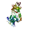
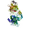
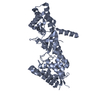
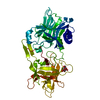
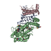
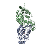
 Tetanospasmin
  Keywords
 Function and homology information
 Authors
 Citation
 Structure visualization
 Downloads & links
 Other downloads

 PDBj
PDBj

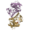
 Assembly
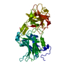

 Mass: 196.967 Da / Num. of mol.: 2 / Source method: obtained synthetically / Formula: Au
 Mass: 18.015 Da / Num. of mol.: 424 / Source method: isolated from a natural source / Formula: H2O
 Sample preparation
 / Beamline: BL1-5 / Wavelength: 1.0396
  Processing