 Movie
Movie Controller

[English] 日本語
 Yorodumi
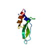
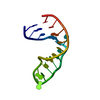
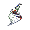
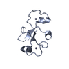
- PDB-1a2e: CRYSTAL STRUCTURE OF A DOUBLE-STRANDED DNA DECAMER CONTAINING A C... -
+ Open data

- Basic information
| Entry | Database: PDB / ID: 1a2e | ||||||
|---|---|---|---|---|---|---|---|
| Title | CRYSTAL STRUCTURE OF A DOUBLE-STRANDED DNA DECAMER CONTAINING A CISPLATIN INTERSTRAND CROSS-LINK ADDUCT | ||||||
 Components Components |
| ||||||
 Keywords Keywords |  DNA / ANTITUMOR DRUG / CIS-DDP / INTERSTRAND CROSS-LINK DNA / ANTITUMOR DRUG / CIS-DDP / INTERSTRAND CROSS-LINK | ||||||
| Function / homology | Cisplatin / DNA Function and homology information Function and homology information | ||||||
| Method | X-RAY DIFFRACTION / SYNCHROTRON / MAD / Resolution: 1.63 Å | ||||||
 Authors Authors | Coste, F. / Zelwer, C. | ||||||
 Citation Citation | Journal: Nucleic Acids Res. / Year: 1999 Title: Crystal structure of a double-stranded DNA containing a cisplatin interstrand cross-link at 1.63 A resolution: hydration at the platinated site. Authors: Coste, F. / Malinge, J.M. / Serre, L. / Shepard, W. / Roth, M. / Leng, M. / Zelwer, C. | ||||||
| History |
|
- Structure visualization
| Structure viewer | Molecule: MolmilJmol/JSmol |
|---|
- Downloads & links
-Download
| PDBx/mmCIF format | 1a2e.cif.gz | 23.6 KB | Display | PDBx/mmCIF format |
|---|---|---|---|---|
| PDB format | pdb1a2e.ent.gz | 14.7 KB | Display | PDB format |
| PDBx/mmJSON format | 1a2e.json.gz | Tree view | PDBx/mmJSON format | |
| Others |  Other downloads Other downloads |
-Validation report
| Arichive directory | https://data.pdbj.org/pub/pdb/validation_reports/a2/1a2eftp://data.pdbj.org/pub/pdb/validation_reports/a2/1a2e | HTTPS FTP |
|---|
-Related structure data
| Similar structure data |
|---|


-Links
 PDBj
PDBj

- Assembly
| Deposited unit | 
| ||||||||||
|---|---|---|---|---|---|---|---|---|---|---|---|
| 1 |
| ||||||||||
| Unit cell |
|
-Components
| #1: DNA chain | Mass: 2931.917 Da / Num. of mol.: 1 / Source method: obtained synthetically / Details: PT(CPT) CROSSLINKS N7(G5) AND N7(G15) |
|---|---|
| #2: DNA chain | Mass: 3159.079 Da / Num. of mol.: 1 / Source method: obtained synthetically / Details: PT(CPT) CROSSLINKS N7(G5) AND N7(G15) |
| #3: Chemical | ChemComp-CPT / Cisplatin  Mass: 300.045 Da / Num. of mol.: 1 / Source method: obtained synthetically / Formula: Cl2H6N2Pt / Comment: medication, chemotherapy*YM Mass: 300.045 Da / Num. of mol.: 1 / Source method: obtained synthetically / Formula: Cl2H6N2Pt / Comment: medication, chemotherapy*YM |
| #4: Water | ChemComp-HOH / Water Mass: 18.015 Da / Num. of mol.: 92 / Source method: isolated from a natural source / Formula: H2O Mass: 18.015 Da / Num. of mol.: 92 / Source method: isolated from a natural source / Formula: H2O |
-Experimental details
-Experiment
| Experiment | Method: X-RAY DIFFRACTION / Number of used crystals: 1 |
|---|
- Sample preparation
| Crystal | Density Matthews: 2.34 Å3/Da / Density % sol: 56 % | ||||||||||||||||||||||||||||||||||||||||||||||||
|---|---|---|---|---|---|---|---|---|---|---|---|---|---|---|---|---|---|---|---|---|---|---|---|---|---|---|---|---|---|---|---|---|---|---|---|---|---|---|---|---|---|---|---|---|---|---|---|---|---|
| Crystal grow | pH: 6.2 / Details: pH 6.20 | ||||||||||||||||||||||||||||||||||||||||||||||||
| Crystal | *PLUS Density % sol: 56 % | ||||||||||||||||||||||||||||||||||||||||||||||||
| Crystal grow | *PLUS Temperature: 4 ℃ / pH: 6 / Method: vapor diffusion, hanging drop | ||||||||||||||||||||||||||||||||||||||||||||||||
| Components of the solutions | *PLUS
|
-Data collection
| Diffraction | Mean temperature: 100 K |
|---|---|
| Diffraction source | Source: SYNCHROTRON / Site: LURE  / Beamline: DW21B / Wavelength: 1.0711 / Beamline: DW21B / Wavelength: 1.0711 |
| Detector | Type: MARRESEARCH / Detector: IMAGE PLATE / Date: Nov 1, 1997 / Details: MIRRORS |
| Radiation | Monochromator: DOUBLE CRYSTAL / Protocol: SINGLE WAVELENGTH / Monochromatic (M) / Laue (L): M / Scattering type: x-ray |
| Radiation wavelength | Wavelength: 1.0711 Å / Relative weight: 1 |
| Reflection | Resolution: 1.63→46.13 Å / Num. obs: 7353 / % possible obs: 93.1 % / Observed criterion σ(I): 4 / Redundancy: 3.5 % / Rmerge(I) obs: 0.037 / Rsym value: 0.037 / Net I/σ(I): 6 |
| Reflection shell | Resolution: 1.63→1.72 Å / Redundancy: 3.1 % / Rmerge(I) obs: 0.063 / Mean I/σ(I) obs: 12 / Rsym value: 0.049 / % possible all: 91 |
| Reflection | *PLUS Num. obs: 7333 / % possible obs: 97 % / Num. measured all: 60517 |
| Reflection shell | *PLUS Redundancy: 3.1 % / Mean I/σ(I) obs: 11.1 |
- Processing
| Software |
| |||||||||||||||||||||||||||||||||
|---|---|---|---|---|---|---|---|---|---|---|---|---|---|---|---|---|---|---|---|---|---|---|---|---|---|---|---|---|---|---|---|---|---|---|
| Refinement | Method to determine structure: MAD / Resolution: 1.63→20 Å / Num. parameters: 2176 / Num. restraintsaints: 2201 / Cross valid method: FREE R / σ(F): 0 / Stereochemistry target values: NUCLSQ
| |||||||||||||||||||||||||||||||||
| Solvent computation | Solvent model: MOEWS & KRETSINGER, J.MOL.BIOL.91(1973)201-223 | |||||||||||||||||||||||||||||||||
| Refine analyze | Occupancy sum hydrogen: 479.67 | |||||||||||||||||||||||||||||||||
| Refinement step | Cycle: LAST / Resolution: 1.63→20 Å
| |||||||||||||||||||||||||||||||||
| Refine LS restraints |
| |||||||||||||||||||||||||||||||||
| Software | *PLUS Name: SHELXL-96 / Classification: refinement | |||||||||||||||||||||||||||||||||
| Refinement | *PLUS Rfactor Rfree: 0.203 | |||||||||||||||||||||||||||||||||
| Solvent computation | *PLUS | |||||||||||||||||||||||||||||||||
| Displacement parameters | *PLUS | |||||||||||||||||||||||||||||||||
| Refine LS restraints | *PLUS
|
