 Movie
Movie Controller

[English] 日本語
 Yorodumi
- PDB-1a2a: AGKISTROTOXIN, A PHOSPHOLIPASE A2-TYPE PRESYNAPTIC NEUROTOXIN FRO... -
+ Open data

- Basic information
| Entry | Database: PDB / ID: 1a2a | ||||||
|---|---|---|---|---|---|---|---|
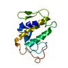
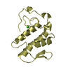
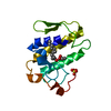
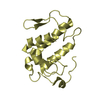
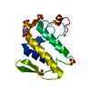
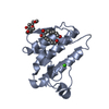
| Title | AGKISTROTOXIN, A PHOSPHOLIPASE A2-TYPE PRESYNAPTIC NEUROTOXIN FROM AGKISTRODON HALYS PALLAS | ||||||
 Components Components | PHOSPHOLIPASE A2 | ||||||
 Keywords Keywords | PRESYNAPTIC NEUROTOXIN / PHOSPHOLIPASE A2 / HYDROLASE / LIPID DEGRADATION | ||||||
| Function / homology |  Function and homology informationphospholipase A2 / phospholipase A2 activity / arachidonic acid secretion / phospholipid metabolic process / lipid catabolic process / toxin activity / calcium ion binding / extracellular region Function and homology informationphospholipase A2 / phospholipase A2 activity / arachidonic acid secretion / phospholipid metabolic process / lipid catabolic process / toxin activity / calcium ion binding / extracellular regionSimilarity search - Function | ||||||
| Biological species |  Gloydius halys (Halys viper) Gloydius halys (Halys viper) | ||||||
| Method | X-RAY DIFFRACTION / MOLECULAR REPLACEMENT / Resolution: 2.8 Å | ||||||
 Authors Authors | Tang, L. / Zhou, Y. / Lin, Z. | ||||||
 Citation Citation | Journal: J.Mol.Biol. / Year: 1998 Title: Crystal structure of agkistrodotoxin, a phospholipase A2-type presynaptic neurotoxin from agkistrodon halys pallas. Authors: Tang, L. / Zhou, Y.C. / Lin, Z.J. #1: Journal: J.Mol.Biol. / Year: 1998Title: Crystal Structure of Agkistrodotoxin, a Phospholipase A2-Type Presynaptic Neurotoxin from Agkistrodon Halys Pallas Authors: Tang, L. / Zhou, Y.C. / Lin, Z.J. | ||||||
| History |
|
- Structure visualization
| Structure viewer | Molecule: MolmilJmol/JSmol |
|---|
- Downloads & links
-Download
| PDBx/mmCIF format | 1a2a.cif.gz | 190.4 KB | Display | PDBx/mmCIF format |
|---|---|---|---|---|
| PDB format | pdb1a2a.ent.gz | 159.1 KB | Display | PDB format |
| PDBx/mmJSON format | 1a2a.json.gz | Tree view | PDBx/mmJSON format | |
| Others |  Other downloads Other downloads |
-Validation report
| Arichive directory | https://data.pdbj.org/pub/pdb/validation_reports/a2/1a2aftp://data.pdbj.org/pub/pdb/validation_reports/a2/1a2a | HTTPS FTP |
|---|
-Related structure data
| Related structure data | 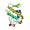 1psjS S: Starting model for refinement |
|---|---|
| Similar structure data |


-Links
 PDBj
PDBj

- Assembly


-Components
| #1: Protein | / AGKISTROTOXIN / ATX Mass: 13870.562 Da / Num. of mol.: 8 / Source method: isolated from a natural source / Source: (natural) Gloydius halys (Halys viper) / References: UniProt: P14421, phospholipase A2#2: Chemical | ChemComp-CL / Chloride  Mass: 35.453 Da / Num. of mol.: 4 / Source method: obtained synthetically / Formula: Cl Mass: 35.453 Da / Num. of mol.: 4 / Source method: obtained synthetically / Formula: Cl |
|---|
-Experimental details
-Experiment
| Experiment | Method: X-RAY DIFFRACTION / Number of used crystals: 1 |
|---|
- Sample preparation
| Crystal | Density Matthews: 2.7 Å3/Da / Density % sol: 49 % | ||||||||||||||||||||||||||||||||||||||||||||||||
|---|---|---|---|---|---|---|---|---|---|---|---|---|---|---|---|---|---|---|---|---|---|---|---|---|---|---|---|---|---|---|---|---|---|---|---|---|---|---|---|---|---|---|---|---|---|---|---|---|---|
| Crystal grow | pH: 4.5 / Details: pH 4.5 | ||||||||||||||||||||||||||||||||||||||||||||||||
| Crystal grow | *PLUS Temperature: 18 ℃ / Method: vapor diffusion, hanging drop | ||||||||||||||||||||||||||||||||||||||||||||||||
| Components of the solutions | *PLUS
|
-Data collection
| Diffraction | Mean temperature: 291 K |
|---|---|
| Diffraction source | Source: ROTATING ANODE / Type: RIGAKU RUH2R / Wavelength: 1.5418 |
| Detector | Type: SIEMENS / Detector: AREA DETECTOR / Date: Sep 1, 1994 / Details: MIRRORS |
| Radiation | Monochromator: NI FILTER / Monochromatic (M) / Laue (L): M / Scattering type: x-ray |
| Radiation wavelength | Wavelength: 1.5418 Å / Relative weight: 1 |
| Reflection | Resolution: 2.58→100 Å / Num. obs: 32147 / % possible obs: 82 % / Observed criterion σ(I): 0 / Redundancy: 1.9 % / Biso Wilson estimate: 28.9 Å2 / Rmerge(I) obs: 0.064 / Rsym value: 0.064 / Net I/σ(I): 9.42 |
| Reflection shell | Resolution: 2.58→2.74 Å / Redundancy: 1.5 % / Rmerge(I) obs: 0.41 / Mean I/σ(I) obs: 1.3 / Rsym value: 0.41 / % possible all: 26 |
| Reflection | *PLUS Highest resolution: 2.59 Å / % possible obs: 84.36 % |
- Processing
| Software |
| ||||||||||||||||||||||||||||||||||||||||||||||||||||||||||||||||||||||||||||||||
|---|---|---|---|---|---|---|---|---|---|---|---|---|---|---|---|---|---|---|---|---|---|---|---|---|---|---|---|---|---|---|---|---|---|---|---|---|---|---|---|---|---|---|---|---|---|---|---|---|---|---|---|---|---|---|---|---|---|---|---|---|---|---|---|---|---|---|---|---|---|---|---|---|---|---|---|---|---|---|---|---|---|
| Refinement | Method to determine structure: MOLECULAR REPLACEMENT Starting model: PDB ENTRY 1PSJ Resolution: 2.8→6 Å / Rfactor Rfree error: 0.008 / Data cutoff high absF: 10000000 / Data cutoff low absF: 0.001 / Isotropic thermal model: RESTRAINED / Cross valid method: THROUGHOUT / σ(F): 2 Details: THE ASYMMETRIC UNIT CONTAINS 8 MOLECULES. THE STRUCTURE WAS SOLVED BY MOLECULAR REPLACEMENT METHOD USING A MONOMERIC SEARCH MODEL WHICH SHOWS 51 PERCENT SEQUENCE IDENTITY.
| ||||||||||||||||||||||||||||||||||||||||||||||||||||||||||||||||||||||||||||||||
| Displacement parameters | Biso mean: 32.7 Å2
| ||||||||||||||||||||||||||||||||||||||||||||||||||||||||||||||||||||||||||||||||
| Refine analyze |
| ||||||||||||||||||||||||||||||||||||||||||||||||||||||||||||||||||||||||||||||||
| Refinement step | Cycle: LAST / Resolution: 2.8→6 Å
| ||||||||||||||||||||||||||||||||||||||||||||||||||||||||||||||||||||||||||||||||
| Refine LS restraints |
| ||||||||||||||||||||||||||||||||||||||||||||||||||||||||||||||||||||||||||||||||
| Refine LS restraints NCS | NCS model details: RESTRAINTS | ||||||||||||||||||||||||||||||||||||||||||||||||||||||||||||||||||||||||||||||||
| LS refinement shell | Resolution: 2.8→2.96 Å / Rfactor Rfree error: 0.03 / Total num. of bins used: 6
| ||||||||||||||||||||||||||||||||||||||||||||||||||||||||||||||||||||||||||||||||
| Xplor file |
| ||||||||||||||||||||||||||||||||||||||||||||||||||||||||||||||||||||||||||||||||
| Software | *PLUS Name: X-PLOR / Version: 3.851 / Classification: refinement | ||||||||||||||||||||||||||||||||||||||||||||||||||||||||||||||||||||||||||||||||
| Refinement | *PLUS % reflection Rfree: 10 % | ||||||||||||||||||||||||||||||||||||||||||||||||||||||||||||||||||||||||||||||||
| Solvent computation | *PLUS | ||||||||||||||||||||||||||||||||||||||||||||||||||||||||||||||||||||||||||||||||
| Displacement parameters | *PLUS | ||||||||||||||||||||||||||||||||||||||||||||||||||||||||||||||||||||||||||||||||
| Refine LS restraints | *PLUS
| ||||||||||||||||||||||||||||||||||||||||||||||||||||||||||||||||||||||||||||||||
| LS refinement shell | *PLUS Rfactor obs: 0.279 |
