 Movie
Movie Controller


+ Open data

- Basic information
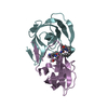
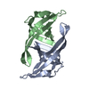
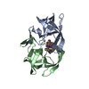
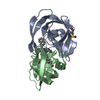
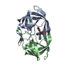
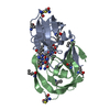
| Entry | Database: PDB / ID: 1a19 | ||||||
|---|---|---|---|---|---|---|---|
| Title | BARSTAR (FREE), C82A MUTANT | ||||||
 Components Components | BARSTAR | ||||||
 Keywords Keywords | RIBONUCLEASE INHIBITOR / BARSTAR / C82A / DIMER / UNCOMPLEXED | ||||||
| Function / homology | Barstar-like / Barstar (barnase inhibitor) / Barstar (barnase inhibitor) / Barstar-like superfamily / Barnase; Chain D / 2-Layer Sandwich / cytoplasm / Alpha Beta / Barstar Function and homology information Function and homology information | ||||||
| Biological species |  Bacillus amyloliquefaciens (bacteria) Bacillus amyloliquefaciens (bacteria) | ||||||
| Method | X-RAY DIFFRACTION / MOLECULAR REPLACEMENT / Resolution: 2.76 Å | ||||||
 Authors Authors | Ratnaparkhi, G.S. / Varadarajan, R. | ||||||
 Citation Citation | Journal: Biochemistry / Year: 1998 Title: Discrepancies between the NMR and X-ray structures of uncomplexed barstar: analysis suggests that packing densities of protein structures determined by NMR are unreliable. Authors: Ratnaparkhi, G.S. / Ramachandran, S. / Udgaonkar, J.B. / Varadarajan, R. | ||||||
| History |
|
- Structure visualization
| Structure viewer | Molecule: MolmilJmol/JSmol |
|---|
- Downloads & links
-Download
| PDBx/mmCIF format | 1a19.cif.gz | 42.7 KB | Display | PDBx/mmCIF format |
|---|---|---|---|---|
| PDB format | pdb1a19.ent.gz | 33.8 KB | Display | PDB format |
| PDBx/mmJSON format | 1a19.json.gz | Tree view | PDBx/mmJSON format | |
| Others |  Other downloads Other downloads |
-Validation report
| Arichive directory | https://data.pdbj.org/pub/pdb/validation_reports/a1/1a19ftp://data.pdbj.org/pub/pdb/validation_reports/a1/1a19 | HTTPS FTP |
|---|
-Related structure data


-Links
 PDBj
PDBj- Assembly
| Deposited unit | 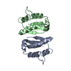
| ||||||||
|---|---|---|---|---|---|---|---|---|---|
| 1 |
| ||||||||
| Unit cell |
|
-Components
| #1: Protein | Mass: 10320.674 Da / Num. of mol.: 2 / Mutation: C82A Source method: isolated from a genetically manipulated source Source: (gene. exp.) Bacillus amyloliquefaciens (bacteria) / Production host: Escherichia coli (E. coli) / References: UniProt: P11540 |
|---|
-Experimental details
-Experiment
| Experiment | Method: X-RAY DIFFRACTION / Number of used crystals: 1 |
|---|
- Sample preparation
| Crystal | Density Matthews: 2.4 Å3/Da / Density % sol: 49 % Description: 1BGS & 2BRS ARE THE COMPLEX OF BARSTAR WITH BARNASE | ||||||||||||||||||||
|---|---|---|---|---|---|---|---|---|---|---|---|---|---|---|---|---|---|---|---|---|---|
| Crystal grow | pH: 6.5 Details: C82A MUTANT WAS CRYSTALLIZED FROM 40-60% AMMONIUM SULFATE, 50MM PHOSPHATE, PH=6.5. PROTEIN CONC=45 MG/ML. C40A AND DTNB LABELLED CRYSTALS ALSO CRYSTALLIZED BUT DID NOT DIFFRACT TO HIGH RESOLUTION. | ||||||||||||||||||||
| Crystal | *PLUS | ||||||||||||||||||||
| Crystal grow | *PLUS Temperature: 20 ℃ / Method: vapor diffusion, hanging drop | ||||||||||||||||||||
| Components of the solutions | *PLUS
|
-Data collection
| Diffraction | Mean temperature: 293 K |
|---|---|
| Diffraction source | Source: ROTATING ANODE / Type: RIGAKU RUH2R / Wavelength: 1.5418 |
| Detector | Type: MARRESEARCH / Detector: IMAGE PLATE / Date: Dec 1, 1996 / Details: MIRRORS |
| Radiation | Monochromatic (M) / Laue (L): M / Scattering type: x-ray |
| Radiation wavelength | Wavelength: 1.5418 Å / Relative weight: 1 |
| Reflection | Resolution: 2.76→10 Å / Num. obs: 4796 / % possible obs: 85 % / Observed criterion σ(I): 2 / Redundancy: 7 % / Biso Wilson estimate: 61.2 Å2 / Rmerge(I) obs: 0.076 / Net I/σ(I): 33 |
| Reflection shell | Resolution: 2.8→2.9 Å / Redundancy: 7 % / Rmerge(I) obs: 0.05 / % possible all: 50 |
| Reflection | *PLUS % possible obs: 99 % / Observed criterion σ(I): 0 |
- Processing
| Software |
| ||||||||||||||||||||||||||||||||||||||||||||||||||||||||||||
|---|---|---|---|---|---|---|---|---|---|---|---|---|---|---|---|---|---|---|---|---|---|---|---|---|---|---|---|---|---|---|---|---|---|---|---|---|---|---|---|---|---|---|---|---|---|---|---|---|---|---|---|---|---|---|---|---|---|---|---|---|---|
| Refinement | Method to determine structure: MOLECULAR REPLACEMENT Starting model: PDB ENTRIES 1BGS, 1BRS Resolution: 2.76→10 Å / Rfactor Rfree error: 0.012 / Data cutoff high absF: 10000000 / Data cutoff low absF: 0.01 / Isotropic thermal model: RESTRAINED / Cross valid method: THROUGHOUT / σ(F): 1 Details: MET PRESENT AT N-TERMINUS DURING EXPRESSION IS NOT INCLUDED IN THE SEQUENCE OR THE STRUCTURE.
| ||||||||||||||||||||||||||||||||||||||||||||||||||||||||||||
| Displacement parameters | Biso mean: 48.4 Å2 | ||||||||||||||||||||||||||||||||||||||||||||||||||||||||||||
| Refine analyze |
| ||||||||||||||||||||||||||||||||||||||||||||||||||||||||||||
| Refinement step | Cycle: LAST / Resolution: 2.76→10 Å
| ||||||||||||||||||||||||||||||||||||||||||||||||||||||||||||
| Refine LS restraints |
| ||||||||||||||||||||||||||||||||||||||||||||||||||||||||||||
| Refine LS restraints NCS | NCS model details: RESTRAINED, 100 KCAL/MOL WEIGHT FINAL POSITIONAL REFINEMENT USING ALL REFLECTIONS AND REMOVING NCS RESTRAINTS. | ||||||||||||||||||||||||||||||||||||||||||||||||||||||||||||
| LS refinement shell | Resolution: 2.8→2.9 Å / Rfactor Rfree error: 0.081 / Total num. of bins used: 10
| ||||||||||||||||||||||||||||||||||||||||||||||||||||||||||||
| Xplor file |
| ||||||||||||||||||||||||||||||||||||||||||||||||||||||||||||
| Software | *PLUS Name: X-PLOR / Version: 3.851 / Classification: refinement | ||||||||||||||||||||||||||||||||||||||||||||||||||||||||||||
| Refine LS restraints | *PLUS
|
