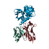
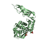
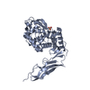
- PDB-1a0i: ATP-DEPENDENT DNA LIGASE FROM BACTERIOPHAGE T7 COMPLEX WITH ATP -
+
Open data
ID or keywords:
Loading...
-
Basic information
Entry
Database: PDB / ID: 1a0i
Title
ATP-DEPENDENT DNA LIGASE FROM BACTERIOPHAGE T7 COMPLEX WITH ATP
Components
DNA LIGASE
Keywords
LIGASE / DNA REPLICATION
Function / homology
Function and homology information
DNA ligation involved in DNA repair / DNA ligase (ATP) / DNA ligase (ATP) activity / double-stranded DNA binding / DNA recombination / DNA replication / ATP binding / metal ion binding Similarity search - Function
DNA ligase, bacteriophage T7-type / DNA ligase, ATP-dependent, bacteriophage T7-type, C-terminal / DNA ligase C-terminal domain / Dna Ligase; domain 1 - #70 / ATP-dependent DNA ligase signature 2. / ATP-dependent DNA ligase AMP-binding site. / DNA ligase/mRNA capping enzyme / DNA ligase, ATP-dependent, conserved site / ATP-dependent DNA ligase family profile. / DNA ligase, ATP-dependent, central ...DNA ligase, bacteriophage T7-type / DNA ligase, ATP-dependent, bacteriophage T7-type, C-terminal / DNA ligase C-terminal domain / Dna Ligase; domain 1 - #70 / ATP-dependent DNA ligase signature 2. / ATP-dependent DNA ligase AMP-binding site. / DNA ligase/mRNA capping enzyme / DNA ligase, ATP-dependent, conserved site / ATP-dependent DNA ligase family profile. / DNA ligase, ATP-dependent, central / ATP dependent DNA ligase domain / D-amino Acid Aminotransferase; Chain A, domain 1 / Nucleic acid-binding proteins / Dna Ligase; domain 1 / OB fold (Dihydrolipoamide Acetyltransferase, E2P) / Nucleic acid-binding, OB-fold / Beta Barrel / 2-Layer Sandwich / Mainly Beta / Alpha Beta Similarity search - Domain/homology
In the structure databanks used in Yorodumi, some data are registered as the other names, "COVID-19 virus" and "2019-nCoV". Here are the details of the virus and the list of structure data.
Jan 31, 2019. EMDB accession codes are about to change! (news from PDBe EMDB page)
EMDB accession codes are about to change! (news from PDBe EMDB page)
The allocation of 4 digits for EMDB accession codes will soon come to an end. Whilst these codes will remain in use, new EMDB accession codes will include an additional digit and will expand incrementally as the available range of codes is exhausted. The current 4-digit format prefixed with “EMD-” (i.e. EMD-XXXX) will advance to a 5-digit format (i.e. EMD-XXXXX), and so on. It is currently estimated that the 4-digit codes will be depleted around Spring 2019, at which point the 5-digit format will come into force.
The EM Navigator/Yorodumi systems omit the EMD- prefix.
Related info.:Q: What is EMD? / ID/Accession-code notation in Yorodumi/EM Navigator
Yorodumi is a browser for structure data from EMDB, PDB, SASBDB, etc.
This page is also the successor to EM Navigator detail page, and also detail information page/front-end page for Omokage search.
The word "yorodu" (or yorozu) is an old Japanese word meaning "ten thousand". "mi" (miru) is to see.
Related info.:EMDB / PDB / SASBDB / Comparison of 3 databanks / Yorodumi Search / Aug 31, 2016. New EM Navigator & Yorodumi / Yorodumi Papers / Jmol/JSmol / Function and homology information / Changes in new EM Navigator and Yorodumi
 Movie
Movie Controller


 Open data
 Basic information
 Components
 Keywords
 Function and homology information

 Authors
 Citation
 Structure visualization
 Downloads & links
 Other downloads
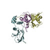


 PDBj
PDBj
 Assembly
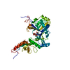


 Mass: 507.181 Da / Num. of mol.: 1 / Source method: obtained synthetically / Formula: C10H16N5O13P3 / Comment: ATP, energy-carrying molecule*YM
 Mass: 18.015 Da / Num. of mol.: 193 / Source method: isolated from a natural source / Formula: H2O
 Sample preparation
 / Beamline: PX7.2 / Wavelength: 1.488
  Processing