 Movie
Movie Controller

[English] 日本語
 Yorodumi
- PDB-1a0h: THE X-RAY CRYSTAL STRUCTURE OF PPACK-MEIZOTHROMBIN DESF1: KRINGLE... -
+ Open data

- Basic information
| Entry | Database: PDB / ID: 1a0h | |||||||||
|---|---|---|---|---|---|---|---|---|---|---|
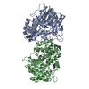
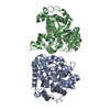
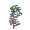
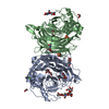
| Title | THE X-RAY CRYSTAL STRUCTURE OF PPACK-MEIZOTHROMBIN DESF1: KRINGLE/THROMBIN AND CARBOHYDRATE/KRINGLE/THROMBIN INTERACTIONS AND LOCATION OF THE LINKER CHAIN | |||||||||
 Components Components | (MEIZOTHROMBIN) x 2 | |||||||||
 Keywords Keywords | HYDROLASE/HYDROLASE INHIBITOR /  SERINE PROTEASE / COAGULATION / THROMBIN / PROTHROMBIN / MEIZOTHROMBIN / HYDROLASE-HYDROLASE INHIBITOR COMPLEX SERINE PROTEASE / COAGULATION / THROMBIN / PROTHROMBIN / MEIZOTHROMBIN / HYDROLASE-HYDROLASE INHIBITOR COMPLEX | |||||||||
| Function / homology |  Function and homology informationfibrinogen binding / thrombin / protein polymerization / positive regulation of blood coagulation / acute-phase response / platelet activation / collagen-containing extracellular matrix / serine-type endopeptidase activity / calcium ion binding / proteolysis / extracellular space Function and homology informationfibrinogen binding / thrombin / protein polymerization / positive regulation of blood coagulation / acute-phase response / platelet activation / collagen-containing extracellular matrix / serine-type endopeptidase activity / calcium ion binding / proteolysis / extracellular spaceSimilarity search - Function | |||||||||
| Biological species |  Bos taurus (cattle) Bos taurus (cattle) | |||||||||
| Method | X-RAY DIFFRACTION / MOLECULAR REPLACEMENT / Resolution: 3.2 Å | |||||||||
 Authors Authors | Martin, P.D. / Malkowski, M.G. / Box, J. / Esmon, C.T. / Edwards, B.F.P. | |||||||||
 Citation Citation | Journal: Structure / Year: 1997 Title: New insights into the regulation of the blood clotting cascade derived from the X-ray crystal structure of bovine meizothrombin des F1 in complex with PPACK. Authors: Martin, P.D. / Malkowski, M.G. / Box, J. / Esmon, C.T. / Edwards, B.F. | |||||||||
| History |
|
- Structure visualization
| Structure viewer | Molecule: MolmilJmol/JSmol |
|---|
- Downloads & links
-Download
| PDBx/mmCIF format | 1a0h.cif.gz | 180.4 KB | Display | PDBx/mmCIF format |
|---|---|---|---|---|
| PDB format | pdb1a0h.ent.gz | 143.1 KB | Display | PDB format |
| PDBx/mmJSON format | 1a0h.json.gz | Tree view | PDBx/mmJSON format | |
| Others |  Other downloads Other downloads |
-Validation report
| Arichive directory | https://data.pdbj.org/pub/pdb/validation_reports/a0/1a0hftp://data.pdbj.org/pub/pdb/validation_reports/a0/1a0h | HTTPS FTP |
|---|
-Related structure data
| Similar structure data |
|---|

-Links
 PDBj
PDBj


- Assembly
| Deposited unit | 
| ||||||||
|---|---|---|---|---|---|---|---|---|---|
| 1 |
| ||||||||
| Unit cell |
| ||||||||
| Components on special symmetry positions |
| ||||||||
| Noncrystallographic symmetry (NCS) | NCS oper: (Code: given Matrix: (-0.90024, -0.43536, -0.00555), Vector : |
-Components
| #1: Protein | Mass: 17868.338 Da / Num. of mol.: 2 / Fragment: F2/THROMBIN DOMAIN / Source method: isolated from a natural source / Source: (natural) Bos taurus (cattle) / Organ: BLOOD / Tissue: BLOOD PLASMA / References: UniProt: P00735, thrombin#2: Protein | Mass: 29772.422 Da / Num. of mol.: 2 / Fragment: F2/THROMBIN DOMAIN / Source method: isolated from a natural source / Source: (natural) Bos taurus (cattle) / Organ: BLOOD / Tissue: BLOOD PLASMA / References: UniProt: P00735, thrombin#3: Polysaccharide | / Mass: 424.401 Da / Num. of mol.: 2Source method: isolated from a genetically manipulated source #4: Chemical |   Type: peptide-like, Peptide-like / Class: Inhibitor / Mass: 453.986 Da / Num. of mol.: 2 / Source method: obtained synthetically / Formula: C21H34ClN6O3 / References: D-Phe-Pro-Arg-CH2Cl Type: peptide-like, Peptide-like / Class: Inhibitor / Mass: 453.986 Da / Num. of mol.: 2 / Source method: obtained synthetically / Formula: C21H34ClN6O3 / References: D-Phe-Pro-Arg-CH2Cl#5: Water | ChemComp-HOH / | Water Mass: 18.015 Da / Num. of mol.: 87 / Source method: isolated from a natural source / Formula: H2O Mass: 18.015 Da / Num. of mol.: 87 / Source method: isolated from a natural source / Formula: H2ONonpolymer details | THE UNBOUND FORM OF THE INHIBITOR IS D-PHE-PRO-ARG-CHLOROMETHYLKETONE. UPON REACTION WITH PROTEIN ...THE UNBOUND FORM OF THE INHIBITOR IS D-PHE-PRO-ARG-CHLOROMETH | |
|---|
-Experimental details
-Experiment
| Experiment | Method: X-RAY DIFFRACTION / Number of used crystals: 1 |
|---|
- Sample preparation
| Crystal | Density Matthews: 5.4 Å3/Da / Density % sol: 76 % | |||||||||||||||||||||||||||||||||||||||||||||||||
|---|---|---|---|---|---|---|---|---|---|---|---|---|---|---|---|---|---|---|---|---|---|---|---|---|---|---|---|---|---|---|---|---|---|---|---|---|---|---|---|---|---|---|---|---|---|---|---|---|---|---|
| Crystal grow | pH: 8 Details: 17 MG/ML PROTEIN, 2% PEG4000, .25 M AMMONIUM PHOSPHATE, PH 8.0, 33% SATURATED AMMONIUM SULFATE | |||||||||||||||||||||||||||||||||||||||||||||||||
| Crystal grow | *PLUS Method: vapor diffusion, hanging drop | |||||||||||||||||||||||||||||||||||||||||||||||||
| Components of the solutions | *PLUS
|
-Data collection
| Diffraction | Mean temperature: 273 K |
|---|---|
| Diffraction source | Source: ROTATING ANODE / Type: RIGAKU RUH2R / Wavelength: 1.5418 |
| Detector | Type: SIEMENS / Detector: AREA DETECTOR / Date: Mar 1, 1996 / Details: 0.3 MM COLLIMATOR |
| Radiation | Monochromator: GRAPHITE(002) / Monochromatic (M) / Laue (L): M / Scattering type: x-ray |
| Radiation wavelength | Wavelength: 1.5418 Å / Relative weight: 1 |
| Reflection | Resolution: 3.2→42.3 Å / Num. obs: 31648 / % possible obs: 88 % / Observed criterion σ(I): 0.5 / Rmerge(I) obs: 0.12 / Rsym value: 0.12 / Net I/σ(I): 5.7 |
| Reflection shell | Resolution: 3.2→3.3 Å / Rmerge(I) obs: 0.19 / Mean I/σ(I) obs: 1.8 / Rsym value: 0.19 / % possible all: 60 |
| Reflection shell | *PLUS % possible obs: 60 % |
- Processing
| Software |
| ||||||||||||||||||||||||
|---|---|---|---|---|---|---|---|---|---|---|---|---|---|---|---|---|---|---|---|---|---|---|---|---|---|
| Refinement | Method to determine structure: MOLECULAR REPLACEMENT Starting model: THROMBIN Resolution: 3.2→7 Å / Data cutoff high absF: 99999999 / Data cutoff low absF: 0.0001 / Cross valid method: THROUGHOUT / σ(F): 1
| ||||||||||||||||||||||||
| Refinement step | Cycle: LAST / Resolution: 3.2→7 Å
| ||||||||||||||||||||||||
| Software | *PLUS Name: X-PLOR / Version: 3.84 / Classification: refinement | ||||||||||||||||||||||||
| Refinement | *PLUS | ||||||||||||||||||||||||
| Solvent computation | *PLUS | ||||||||||||||||||||||||
| Displacement parameters | *PLUS | ||||||||||||||||||||||||
| Refine LS restraints | *PLUS
|
