 Movie
Movie Controller

[English] 日本語
 Yorodumi
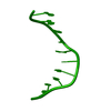
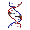
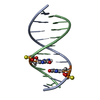
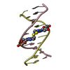
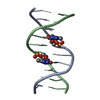
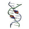
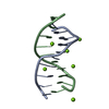
- PDB-183d: X-RAY STRUCTURE OF A DNA DECAMER CONTAINING 7, 8-DIHYDRO-8-OXOGUANINE -
+ Open data

- Basic information
| Entry | Database: PDB / ID: 183d | ||||||||||||||||||
|---|---|---|---|---|---|---|---|---|---|---|---|---|---|---|---|---|---|---|---|
| Title | X-RAY STRUCTURE OF A DNA DECAMER CONTAINING 7, 8-DIHYDRO-8-OXOGUANINE | ||||||||||||||||||
 Components Components | DNA (5'-D(* Keywords Keywords DNA / B-DNA / DOUBLE HELIX / MODIFIED DNA / B-DNA / DOUBLE HELIX / MODIFIEDFunction / homology | DNA Function and homology information Function and homology informationMethod | X-RAY DIFFRACTION / Resolution: 1.6 Å  Authors AuthorsLipscomb, L.A. / Peek, M.E. / Morningstar, M.L. / Verghis, S.M. / Miller, E.M. / Rich, A. / Essigmann, J.M. / Williams, L.D. |  CitationJournal: Proc.Natl.Acad.Sci.USA / Year: 1995 CitationJournal: Proc.Natl.Acad.Sci.USA / Year: 1995Title: X-ray structure of a DNA decamer containing 7,8-dihydro-8-oxoguanine. Authors: Lipscomb, L.A. / Peek, M.E. / Morningstar, M.L. / Verghis, S.M. / Miller, E.M. / Rich, A. / Essigmann, J.M. / Williams, L.D. History |
|
- Structure visualization
| Structure viewer | Molecule: MolmilJmol/JSmol |
|---|
- Downloads & links
-Download
| PDBx/mmCIF format | 183d.cif.gz | 15.9 KB | Display | PDBx/mmCIF format |
|---|---|---|---|---|
| PDB format | pdb183d.ent.gz | 9.1 KB | Display | PDB format |
| PDBx/mmJSON format | 183d.json.gz | Tree view | PDBx/mmJSON format | |
| Others |  Other downloads Other downloads |
-Validation report
| Arichive directory | https://data.pdbj.org/pub/pdb/validation_reports/83/183dftp://data.pdbj.org/pub/pdb/validation_reports/83/183d | HTTPS FTP |
|---|
-Related structure data
| Similar structure data |
|---|


-Links
 PDBj
PDBj

- Assembly
| Deposited unit | 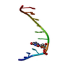
| ||||||||
|---|---|---|---|---|---|---|---|---|---|
| 1 | 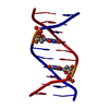
| ||||||||
| Unit cell |
|
-Components
| #1: DNA chain | Mass: 3061.992 Da / Num. of mol.: 1 / Source method: obtained synthetically |
|---|---|
| #2: Water | ChemComp-HOH / Water Mass: 18.015 Da / Num. of mol.: 42 / Source method: isolated from a natural source / Formula: H2O Mass: 18.015 Da / Num. of mol.: 42 / Source method: isolated from a natural source / Formula: H2O |
-Experimental details
-Experiment
| Experiment | Method: X-RAY DIFFRACTION |
|---|
- Sample preparation
| Crystal | Density Matthews: 2.11 Å3/Da / Density % sol: 41.57 % | |||||||||||||||||||||||||||||||||||||||||||||||||
|---|---|---|---|---|---|---|---|---|---|---|---|---|---|---|---|---|---|---|---|---|---|---|---|---|---|---|---|---|---|---|---|---|---|---|---|---|---|---|---|---|---|---|---|---|---|---|---|---|---|---|
| Crystal grow | Temperature: 277 K / Method: vapor diffusion, sitting drop / pH: 7.6 Details: pH 7.60, VAPOR DIFFUSION, SITTING DROP, temperature 277.00K | |||||||||||||||||||||||||||||||||||||||||||||||||
| Components of the solutions |
| |||||||||||||||||||||||||||||||||||||||||||||||||
| Crystal grow | *PLUS Temperature: 4 ℃ / pH: 7.6 | |||||||||||||||||||||||||||||||||||||||||||||||||
| Components of the solutions | *PLUS
|
-Data collection
| Diffraction | Mean temperature: 277 K |
|---|---|
| Diffraction source | Source: ROTATING ANODE / Type: RIGAKU RU200 |
| Detector | Type: SDMS / Detector: AREA DETECTOR |
| Radiation | Scattering type: x-ray |
| Radiation wavelength | Relative weight: 1 |
| Reflection | Highest resolution: 1.6 Å / Num. all: 12930 / Num. obs: 3290 / Observed criterion σ(I): 2 |
| Reflection | *PLUS Highest resolution: 1.6 Å / Observed criterion σ(I): 2 / Rmerge(I) obs: 0.0552 |
- Processing
| Software | Name: X-PLOR / Classification: refinement | ||||||||||||||||||||||||||||||||||||||||||||||||||||||||||||
|---|---|---|---|---|---|---|---|---|---|---|---|---|---|---|---|---|---|---|---|---|---|---|---|---|---|---|---|---|---|---|---|---|---|---|---|---|---|---|---|---|---|---|---|---|---|---|---|---|---|---|---|---|---|---|---|---|---|---|---|---|---|
| Refinement | Resolution: 1.6→10 Å / σ(F): 2
| ||||||||||||||||||||||||||||||||||||||||||||||||||||||||||||
| Refine Biso |
| ||||||||||||||||||||||||||||||||||||||||||||||||||||||||||||
| Refinement step | Cycle: LAST / Resolution: 1.6→10 Å
| ||||||||||||||||||||||||||||||||||||||||||||||||||||||||||||
| Refine LS restraints |
| ||||||||||||||||||||||||||||||||||||||||||||||||||||||||||||
| Refinement | *PLUS Highest resolution: 1.6 Å / Lowest resolution: 10 Å / σ(F): 2 | ||||||||||||||||||||||||||||||||||||||||||||||||||||||||||||
| Solvent computation | *PLUS | ||||||||||||||||||||||||||||||||||||||||||||||||||||||||||||
| Displacement parameters | *PLUS | ||||||||||||||||||||||||||||||||||||||||||||||||||||||||||||
| Refine LS restraints | *PLUS Type: x_angle_deg / Dev ideal: 3.6 |
