 Movie
Movie Controller

[English] 日本語
 Yorodumi
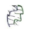
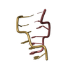
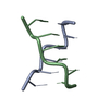
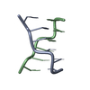
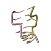
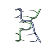
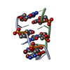
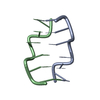
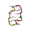
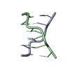
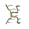
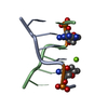
- PDB-181d: SEQUENCE-DEPENDENT MICROHETEROGENEITY OF Z-DNA: THE CRYSTAL AND M... -
+ Open data

- Basic information
| Entry | Database: PDB / ID: 181d | ||||||
|---|---|---|---|---|---|---|---|
| Title | SEQUENCE-DEPENDENT MICROHETEROGENEITY OF Z-DNA: THE CRYSTAL AND MOLECULAR STRUCTURES OF D(CACGCG).D(CGCGTG) AND D(CGCACG).D(CGTGCG) | ||||||
 Components Components |
| ||||||
 Keywords Keywords |  DNA / Z-DNA / DOUBLE HELIX DNA / Z-DNA / DOUBLE HELIX | ||||||
| Function / homology | DNA Function and homology information Function and homology information | ||||||
| Method | X-RAY DIFFRACTION / Resolution: 1.6 Å | ||||||
 Authors Authors | Sadasivan, C. / Gautham, N. | ||||||
 Citation Citation | Journal: J.Mol.Biol. / Year: 1995 Title: Sequence-dependent microheterogeneity of Z-DNA: the crystal and molecular structures of d(CACGCG).d(CGCGTG) and d(CGCACG).d(CGTGCG). Authors: Sadasivan, C. / Gautham, N. #1: Journal: To be PublishedTitle: Plasticity of Z-DNA as Observed in the Crystal Structures of Non-Selfcomplementary Hexanucleotides Authors: Sadasivan, C. / Gautham, N. #2: Journal: Acta Crystallogr.,Sect.D / Year: 1994Title: Space Group Degeneracy in the Packing of a Non-Selfcomplementary Z-DNA Hexamer Authors: Sadasivan, C. / Karthe, P. / Gautham, N. | ||||||
| History |
|
- Structure visualization
| Structure viewer | Molecule: MolmilJmol/JSmol |
|---|
- Downloads & links
-Download
| PDBx/mmCIF format | 181d.cif.gz | 17 KB | Display | PDBx/mmCIF format |
|---|---|---|---|---|
| PDB format | pdb181d.ent.gz | 9.8 KB | Display | PDB format |
| PDBx/mmJSON format | 181d.json.gz | Tree view | PDBx/mmJSON format | |
| Others |  Other downloads Other downloads |
-Validation report
| Arichive directory | https://data.pdbj.org/pub/pdb/validation_reports/81/181dftp://data.pdbj.org/pub/pdb/validation_reports/81/181d | HTTPS FTP |
|---|
-Related structure data


-Links
 PDBj
PDBj

- Assembly
| Deposited unit | 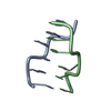
| ||||||||
|---|---|---|---|---|---|---|---|---|---|
| 1 |
| ||||||||
| Unit cell |
|
-Components
| #1: DNA chain | Mass: 1794.206 Da / Num. of mol.: 1 / Source method: obtained synthetically |
|---|---|
| #2: DNA chain | Mass: 1825.216 Da / Num. of mol.: 1 / Source method: obtained synthetically |
| #3: Water | ChemComp-HOH / Water Mass: 18.015 Da / Num. of mol.: 42 / Source method: isolated from a natural source / Formula: H2O Mass: 18.015 Da / Num. of mol.: 42 / Source method: isolated from a natural source / Formula: H2O |
-Experimental details
-Experiment
| Experiment | Method: X-RAY DIFFRACTION |
|---|
- Sample preparation
| Crystal | Density Matthews: 1.7 Å3/Da / Density % sol: 27.63 % | |||||||||||||||||||||||||||||||||||
|---|---|---|---|---|---|---|---|---|---|---|---|---|---|---|---|---|---|---|---|---|---|---|---|---|---|---|---|---|---|---|---|---|---|---|---|---|
| Crystal grow | Method: vapor diffusion, hanging drop / pH: 6.9 / Details: pH 6.90, VAPOR DIFFUSION, HANGING DROP / Temp details: ROOM TEMPERATURE | |||||||||||||||||||||||||||||||||||
| Components of the solutions |
| |||||||||||||||||||||||||||||||||||
| Crystal grow | *PLUS pH: 6.9 | |||||||||||||||||||||||||||||||||||
| Components of the solutions | *PLUS
|
-Data collection
| Diffraction | Mean temperature: 296 K |
|---|---|
| Detector | Type: ENRAF-NONIUS CAD4 / Detector: DIFFRACTOMETER |
| Radiation | Scattering type: x-ray |
| Radiation wavelength | Relative weight: 1 |
| Reflection | Highest resolution: 1.6 Å / Num. obs: 3912 |
| Reflection | *PLUS Highest resolution: 1.6 Å |
- Processing
| Software | Name: NUCLSQ / Classification: refinement | ||||||||||||||||||||||||||||||||||||||||||||||||||||||||||||||||||||||||||||||||||||
|---|---|---|---|---|---|---|---|---|---|---|---|---|---|---|---|---|---|---|---|---|---|---|---|---|---|---|---|---|---|---|---|---|---|---|---|---|---|---|---|---|---|---|---|---|---|---|---|---|---|---|---|---|---|---|---|---|---|---|---|---|---|---|---|---|---|---|---|---|---|---|---|---|---|---|---|---|---|---|---|---|---|---|---|---|---|
| Refinement | Resolution: 1.6→8 Å / σ(F): 2 /
| ||||||||||||||||||||||||||||||||||||||||||||||||||||||||||||||||||||||||||||||||||||
| Refine Biso |
| ||||||||||||||||||||||||||||||||||||||||||||||||||||||||||||||||||||||||||||||||||||
| Refinement step | Cycle: LAST / Resolution: 1.6→8 Å
| ||||||||||||||||||||||||||||||||||||||||||||||||||||||||||||||||||||||||||||||||||||
| Refine LS restraints |
| ||||||||||||||||||||||||||||||||||||||||||||||||||||||||||||||||||||||||||||||||||||
| Refinement | *PLUS Highest resolution: 1.6 Å / Lowest resolution: 8 Å / σ(F): 2 | ||||||||||||||||||||||||||||||||||||||||||||||||||||||||||||||||||||||||||||||||||||
| Solvent computation | *PLUS | ||||||||||||||||||||||||||||||||||||||||||||||||||||||||||||||||||||||||||||||||||||
| Displacement parameters | *PLUS |
