- PDB-173d: MULTIPLE BINDING MODES OF ANTICANCER DRUG ACTINOMYCIN D: X-RAY, M... -
+
Open data
ID or keywords:
Loading...
-
Basic information
Entry
Database: PDB / ID: 173d
Title
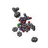
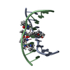
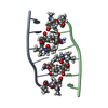
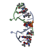
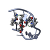
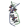
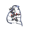
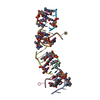
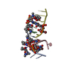
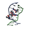
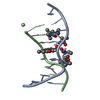
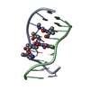
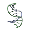
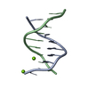
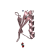
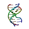
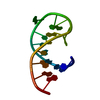
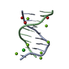
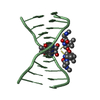
MULTIPLE BINDING MODES OF ANTICANCER DRUG ACTINOMYCIN D: X-RAY, MOLECULAR MODELING, AND SPECTROSCOPIC STUDIES OF D(GAAGCTTC)2-ACTINOMYCIN D COMPLEXES AND ITS HOST DNA
Journal: J.Am.Chem.Soc. / Year: 1994 Title: Multiple Binding Modes of Anticancer Drug Actinomycin D: X-Ray, Molecular Modeling, and Spectroscopic Studies of D(Gaagcttc)2-Actinomycin D Complexes and its Host DNA Authors: Kamitori, S. / Takusagawa, F.
History
Deposition
Apr 18, 1994
Deposition site: BNL / Processing site: NDB
Revision 1.0
Oct 15, 1994
Provider: repository / Type: Initial release
Revision 1.1
Jun 14, 2011
Group: Version format compliance
Revision 1.2
Jul 13, 2011
Group: Version format compliance
Revision 1.3
Jul 27, 2011
Group: Atomic model / Database references ...Atomic model / Database references / Derived calculations / Non-polymer description / Structure summary
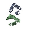
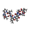
Type: PolypeptidePeptide / Class: Antibiotic / Mass: 1259.447 Da / Num. of mol.: 2 / Source method: isolated from a natural source Details: ACTINOMYCIN D CONSISTS OF TWO PENTAMER RINGS LINKED BY THE CHROMOPHORE (PXZ) Source: (natural) STREPTOMYCES ANTIBIOTICUS (bacteria) / References: NOR: NOR00228, Actinomycin D
Mass: 18.015 Da / Num. of mol.: 96 / Source method: isolated from a natural source / Formula: H2O
Compound details
ACTINOMYCIN D IS A BICYCLIC PEPTIDE, A MEMBER OF THE ACTINOMYCIN FAMILY. HERE, ACTINOMYCIN D IS ...ACTINOMYCIN D IS A BICYCLIC PEPTIDE, A MEMBER OF THE ACTINOMYCIN FAMILY. HERE, ACTINOMYCIN D IS REPRESENTED BY THE SEQUENCE (SEQRES)
-
Experimental details
-
Experiment
Experiment
Method: X-RAY DIFFRACTION
-
Sample preparation
Crystal
Density Matthews: 2.88 Å3/Da / Density % sol: 57.36 %
Crystal grow
pH: 7 / Details: PH 7.00, VAPOR DIFFUSION, TEMPERATURE 277.00K
Components of the solutions
ID
Name
Crystal-ID
Sol-ID
1
WATER
1
1
2
MPD
1
1
3
MGCL2
1
1
4
NACACODYLATE
1
1
5
SPERMINE_HCL
1
1
6
WATER
1
2
7
MPD
1
2
Crystal grow
*PLUS
Temperature: 4 ℃ / pH: 7 / Method: unknown
Components of the solutions
*PLUS
ID
Conc.
Common name
Crystal-ID
Chemical formula
Sol-ID
1
2.0mM
oligonucleotide
1
2
10mM
1
MgCl2
3
20mM
cacodylate
1
4
2.0mM
sperminetetrachloride
1
5
1.5mM
ActinomycinD
1
6
7.5 %(v/v)
MPD
1
1
7
15 %(v/v)
1
-
Data collection
Diffraction
Mean temperature: 123 K
Detector
Type: RIGAKU AFC-5R / Detector: DIFFRACTOMETER
Radiation
Protocol: SINGLE WAVELENGTH / Monochromatic (M) / Laue (L): M / Scattering type: x-ray
Radiation wavelength
Relative weight: 1
Reflection
Highest resolution: 3 Å / Num. obs: 1016
-
Processing
Software
Name: X-PLOR / Classification: refinement
Refinement
Resolution: 3→8 Å / σ(F): 2 /
Rfactor
Num. reflection
Rwork
0.202
-
obs
0.202
1016
Refinement step
Cycle: LAST / Resolution: 3→8 Å
Protein
Nucleic acid
Ligand
Solvent
Total
Num. atoms
94
322
0
96
512
Refine LS restraints
Refine-ID
Type
Dev ideal
X-RAY DIFFRACTION
x_bond_d
0.037
X-RAY DIFFRACTION
x_bond_d_na
X-RAY DIFFRACTION
x_bond_d_prot
X-RAY DIFFRACTION
x_angle_d
X-RAY DIFFRACTION
x_angle_d_na
X-RAY DIFFRACTION
x_angle_d_prot
X-RAY DIFFRACTION
x_angle_deg
5.9
X-RAY DIFFRACTION
x_angle_deg_na
X-RAY DIFFRACTION
x_angle_deg_prot
X-RAY DIFFRACTION
x_dihedral_angle_d
X-RAY DIFFRACTION
x_dihedral_angle_d_na
X-RAY DIFFRACTION
x_dihedral_angle_d_prot
X-RAY DIFFRACTION
x_improper_angle_d
X-RAY DIFFRACTION
x_improper_angle_d_na
X-RAY DIFFRACTION
x_improper_angle_d_prot
X-RAY DIFFRACTION
x_mcbond_it
X-RAY DIFFRACTION
x_mcangle_it
X-RAY DIFFRACTION
x_scbond_it
X-RAY DIFFRACTION
x_scangle_it
Refinement
*PLUS
Highest resolution: 3 Å / Lowest resolution: 8 Å / σ(F): 2 / Rfactor obs: 0.202
Solvent computation
*PLUS
Displacement parameters
*PLUS
Refine LS restraints
*PLUS
Type: x_angle_d / Dev ideal: 5.9
+
About Yorodumi
-
News
-
Feb 9, 2022. New format data for meta-information of EMDB entries
New format data for meta-information of EMDB entries
Version 3 of the EMDB header file is now the official format.
The previous official version 1.9 will be removed from the archive.
In the structure databanks used in Yorodumi, some data are registered as the other names, "COVID-19 virus" and "2019-nCoV". Here are the details of the virus and the list of structure data.
Jan 31, 2019. EMDB accession codes are about to change! (news from PDBe EMDB page)
EMDB accession codes are about to change! (news from PDBe EMDB page)
The allocation of 4 digits for EMDB accession codes will soon come to an end. Whilst these codes will remain in use, new EMDB accession codes will include an additional digit and will expand incrementally as the available range of codes is exhausted. The current 4-digit format prefixed with “EMD-” (i.e. EMD-XXXX) will advance to a 5-digit format (i.e. EMD-XXXXX), and so on. It is currently estimated that the 4-digit codes will be depleted around Spring 2019, at which point the 5-digit format will come into force.
The EM Navigator/Yorodumi systems omit the EMD- prefix.
Related info.:Q: What is EMD? / ID/Accession-code notation in Yorodumi/EM Navigator
Yorodumi is a browser for structure data from EMDB, PDB, SASBDB, etc.
This page is also the successor to EM Navigator detail page, and also detail information page/front-end page for Omokage search.
The word "yorodu" (or yorozu) is an old Japanese word meaning "ten thousand". "mi" (miru) is to see.
Related info.:EMDB / PDB / SASBDB / Comparison of 3 databanks / Yorodumi Search / Aug 31, 2016. New EM Navigator & Yorodumi / Yorodumi Papers / Jmol/JSmol / Function and homology information / Changes in new EM Navigator and Yorodumi
 Movie
Movie Controller

 Yorodumi
 Open data
 Basic information
 Components
 Keywords
 ACTINOMYCIN /
  Function and homology information
 Authors
 Citation
 Structure visualization
 Downloads & links
 Other downloads


 PDBj
PDBj
 Assembly
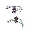

 Type: Polypeptide
 Mass: 18.015 Da / Num. of mol.: 96 / Source method: isolated from a natural source / Formula: H2O
 Sample preparation
 Processing