 Movie
Movie Controller

[English] 日本語
 Yorodumi
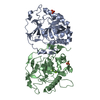
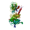
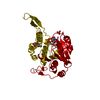
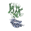
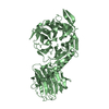
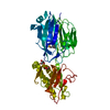
- PDB-117e: THE R78K AND D117E ACTIVE SITE VARIANTS OF SACCHAROMYCES CEREVISI... -
+ Open data

- Basic information
| Entry | Database: PDB / ID: 117e | ||||||
|---|---|---|---|---|---|---|---|
| Title | THE R78K AND D117E ACTIVE SITE VARIANTS OF SACCHAROMYCES CEREVISIAE SOLUBLE INORGANIC PYROPHOSPHATASE: STRUCTURAL STUDIES AND MECHANISTIC IMPLICATIONS | ||||||
 Components Components | PROTEIN (INORGANIC PYROPHOSPHATASE) | ||||||
 Keywords Keywords |  HYDROLASE / ENZYME MECHANISM / IORGANIC PYROPHOSPHATASE / MUTAN STRUCTURES / 2-METAL ION MECHANISM HYDROLASE / ENZYME MECHANISM / IORGANIC PYROPHOSPHATASE / MUTAN STRUCTURES / 2-METAL ION MECHANISM | ||||||
| Function / homology |  Function and homology information Function and homology informationCytosolic tRNA aminoacylation / Mitochondrial tRNA aminoacylation / Pyrophosphate hydrolysis / inorganic diphosphatase / inorganic diphosphate phosphatase activity / phosphate-containing compound metabolic process / magnesium ion binding / nucleus / cytoplasmSimilarity search - Function | ||||||
| Biological species |  Saccharomyces cerevisiae (brewer's yeast) Saccharomyces cerevisiae (brewer's yeast) | ||||||
| Method | X-RAY DIFFRACTION / SYNCHROTRON / MOLECULAR REPLACEMENT / Resolution: 2.15 Å | ||||||
 Authors Authors | Tuominen, V. / Heikinheimo, P. / Kajander, T. / Torkkel, T. / Hyytia, T. / Kapyla, J. / Lahti, R. / Cooperman, B.S. / Goldman, A. | ||||||
 Citation Citation | Journal: J.Mol.Biol. / Year: 1998 Title: The R78K and D117E active-site variants of Saccharomyces cerevisiae soluble inorganic pyrophosphatase: structural studies and mechanistic implications. Authors: Tuominen, V. / Heikinheimo, P. / Kajander, T. / Torkkel, T. / Hyytia, T. / Kapyla, J. / Lahti, R. / Cooperman, B.S. / Goldman, A. | ||||||
| History |
|
- Structure visualization
| Structure viewer | Molecule: MolmilJmol/JSmol |
|---|
- Downloads & links
-Download
| PDBx/mmCIF format | 117e.cif.gz | 134.1 KB | Display | PDBx/mmCIF format |
|---|---|---|---|---|
| PDB format | pdb117e.ent.gz | 102.3 KB | Display | PDB format |
| PDBx/mmJSON format | 117e.json.gz | Tree view | PDBx/mmJSON format | |
| Others |  Other downloads Other downloads |
-Validation report
| Arichive directory | https://data.pdbj.org/pub/pdb/validation_reports/17/117eftp://data.pdbj.org/pub/pdb/validation_reports/17/117e | HTTPS FTP |
|---|
-Related structure data
| Related structure data |  8prkC 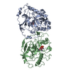 1wgjS S: Starting model for refinement C: citing same article ( |
|---|---|
| Similar structure data |


-Links
 PDBj
PDBj- Assembly
| Deposited unit | 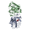
| ||||||||
|---|---|---|---|---|---|---|---|---|---|
| 1 |
| ||||||||
| Unit cell |
| ||||||||
| Noncrystallographic symmetry (NCS) | NCS oper: (Code: given Matrix: (0.798845, 0.601385, 0.013493), Vector : |
-Components
| #1: Protein | Mass: 32239.398 Da / Num. of mol.: 2 / Mutation: D117E Source method: isolated from a genetically manipulated source Details: PRODUCT COMPLEX Source: (gene. exp.) Saccharomyces cerevisiae (brewer's yeast)Cellular location: CYTOPLASM / Gene: PPA1 / Production host:  Escherichia coli (E. coli) / Strain (production host): HB101 / References: UniProt: P00817, inorganic diphosphatase Escherichia coli (E. coli) / Strain (production host): HB101 / References: UniProt: P00817, inorganic diphosphatase#2: Chemical | ChemComp-MN /   Mass: 54.938 Da / Num. of mol.: 8 / Source method: obtained synthetically / Formula: Mn Mass: 54.938 Da / Num. of mol.: 8 / Source method: obtained synthetically / Formula: Mn#3: Chemical | Phosphate  Mass: 94.971 Da / Num. of mol.: 3 / Source method: obtained synthetically / Formula: PO4 Mass: 94.971 Da / Num. of mol.: 3 / Source method: obtained synthetically / Formula: PO4#4: Water | ChemComp-HOH / | Water Mass: 18.015 Da / Num. of mol.: 454 / Source method: isolated from a natural source / Formula: H2O Mass: 18.015 Da / Num. of mol.: 454 / Source method: isolated from a natural source / Formula: H2O |
|---|
-Experimental details
-Experiment
| Experiment | Method: X-RAY DIFFRACTION / Number of used crystals: 1 |
|---|
- Sample preparation
| Crystal | Density Matthews: 2.71 Å3/Da / Density % sol: 54.69 % | ||||||||||||||||||||||||||||||||||||||||||
|---|---|---|---|---|---|---|---|---|---|---|---|---|---|---|---|---|---|---|---|---|---|---|---|---|---|---|---|---|---|---|---|---|---|---|---|---|---|---|---|---|---|---|---|
| Crystal grow | pH: 6 Details: 17-19% MPD, 25 MM MES, PH 6.0, 1 MM MNCL2, 0.5 MM NA2HPO4, 10 MG/ML PROTEIN | ||||||||||||||||||||||||||||||||||||||||||
| Crystal grow | *PLUS Temperature: 4 ℃ / Method: vapor diffusion, sitting drop | ||||||||||||||||||||||||||||||||||||||||||
| Components of the solutions | *PLUS
|
-Data collection
| Diffraction | Mean temperature: 100 K |
|---|---|
| Diffraction source | Source: SYNCHROTRON / Site: EMBL/DESY, HAMBURG  / Beamline: BW7A / Wavelength: 0.891 / Beamline: BW7A / Wavelength: 0.891 |
| Detector | Type: MARRESEARCH / Detector: IMAGE PLATE / Date: Sep 1, 1996 |
| Radiation | Protocol: SINGLE WAVELENGTH / Monochromatic (M) / Laue (L): M / Scattering type: x-ray |
| Radiation wavelength | Wavelength: 0.891 Å / Relative weight: 1 |
| Reflection | Resolution: 2.15→20 Å / Num. obs: 36385 / % possible obs: 88.9 % / Observed criterion σ(I): 2 / Redundancy: 6.3 % / Biso Wilson estimate: 2.7 Å2 / Rmerge(I) obs: 0.086 / Net I/σ(I): 5.7 |
| Reflection shell | Resolution: 2.15→2.19 Å / % possible all: 86 |
| Reflection | *PLUS Num. measured all: 228918 |
| Reflection shell | *PLUS % possible obs: 86 % |
- Processing
| Software |
| ||||||||||||||||||||||||||||||||||||||||||||||||||||||||||||||||||||||||||||||||
|---|---|---|---|---|---|---|---|---|---|---|---|---|---|---|---|---|---|---|---|---|---|---|---|---|---|---|---|---|---|---|---|---|---|---|---|---|---|---|---|---|---|---|---|---|---|---|---|---|---|---|---|---|---|---|---|---|---|---|---|---|---|---|---|---|---|---|---|---|---|---|---|---|---|---|---|---|---|---|---|---|---|
| Refinement | Method to determine structure: MOLECULAR REPLACEMENT Starting model: 1WGJ Resolution: 2.15→8 Å / Rfactor Rfree error: 0.006 / Data cutoff high absF: 10000000 / Data cutoff low absF: 0.001 / Isotropic thermal model: RESTRAINED / Cross valid method: THROUGHOUT / σ(F): 2 / Details: BULK SOLVENT MODEL USED
| ||||||||||||||||||||||||||||||||||||||||||||||||||||||||||||||||||||||||||||||||
| Displacement parameters | Biso mean: 9.7 Å2
| ||||||||||||||||||||||||||||||||||||||||||||||||||||||||||||||||||||||||||||||||
| Refine analyze |
| ||||||||||||||||||||||||||||||||||||||||||||||||||||||||||||||||||||||||||||||||
| Refinement step | Cycle: LAST / Resolution: 2.15→8 Å
| ||||||||||||||||||||||||||||||||||||||||||||||||||||||||||||||||||||||||||||||||
| Refine LS restraints |
| ||||||||||||||||||||||||||||||||||||||||||||||||||||||||||||||||||||||||||||||||
| LS refinement shell | Resolution: 2.15→2.19 Å / Rfactor Rfree error: 0.03 / Total num. of bins used: 20
| ||||||||||||||||||||||||||||||||||||||||||||||||||||||||||||||||||||||||||||||||
| Xplor file |
| ||||||||||||||||||||||||||||||||||||||||||||||||||||||||||||||||||||||||||||||||
| Software | *PLUS Name: X-PLOR / Version: 3.851 / Classification: refinement | ||||||||||||||||||||||||||||||||||||||||||||||||||||||||||||||||||||||||||||||||
| Refine LS restraints | *PLUS
|
