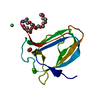
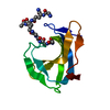
 Movie
Movie Controller


+ Open data

- Basic information
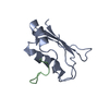
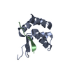
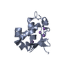
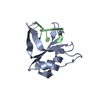
| Entry | Database: PDB / ID: 5hdg | ||||||
|---|---|---|---|---|---|---|---|
| Title | crystal structure of heat shock factor 1-DBD | ||||||
 Components Components | Heat shock factor protein 1 | ||||||
 Keywords Keywords | TRANSCRIPTION / SF1-DBD | ||||||
| Function / homology |  Function and homology information Function and homology informationcellular response to nitroglycerin / response to hypobaric hypoxia / sequence-specific single stranded DNA binding / cellular response to L-glutamine / cellular response to diamide / negative regulation of double-strand break repair via nonhomologous end joining / cellular response to sodium arsenite / positive regulation of apoptotic DNA fragmentation / translation elongation factor binding / negative regulation of inclusion body assembly ...cellular response to nitroglycerin / response to hypobaric hypoxia / sequence-specific single stranded DNA binding / cellular response to L-glutamine / cellular response to diamide / negative regulation of double-strand break repair via nonhomologous end joining / cellular response to sodium arsenite / positive regulation of apoptotic DNA fragmentation / translation elongation factor binding / negative regulation of inclusion body assembly / positive regulation of inclusion body assembly / nuclear stress granule / cellular response to potassium ion / positive regulation of macrophage differentiation / protein folding chaperone complex / cellular response to angiotensin / negative regulation of cardiac muscle cell apoptotic process / response to psychosocial stress / STAT family protein binding / RNA polymerase II intronic transcription regulatory region sequence-specific DNA binding / response to testosterone / mitotic spindle pole / general transcription initiation factor binding / HSF1-dependent transactivation / Regulation of HSF1-mediated heat shock response / HSF1 activation / mRNA transport / Attenuation phase / cellular response to unfolded protein / negative regulation of protein-containing complex assembly / heterochromatin / regulation of cellular response to heat / positive regulation of tyrosine phosphorylation of STAT protein / cellular response to copper ion / cellular response to cadmium ion / heat shock protein binding / positive regulation of mitotic cell cycle / response to nutrient / response to activity / cellular response to estradiol stimulus / promoter-specific chromatin binding / Hsp90 protein binding / euchromatin / cellular response to gamma radiation / defense response / PML body / chromatin DNA binding / kinetochore / cellular response to hydrogen peroxide / mRNA processing / DNA-binding transcription repressor activity, RNA polymerase II-specific / Aggrephagy / : / positive regulation of DNA-binding transcription factor activity / MAPK cascade / cellular response to xenobiotic stimulus / sequence-specific double-stranded DNA binding / cellular response to heat / positive regulation of cold-induced thermogenesis / DNA-binding transcription activator activity, RNA polymerase II-specific / protein-containing complex assembly / cellular response to lipopolysaccharide / sequence-specific DNA binding / transcription cis-regulatory region binding / DNA-binding transcription factor activity, RNA polymerase II-specific / ribonucleoprotein complex / RNA polymerase II cis-regulatory region sequence-specific DNA binding / DNA-binding transcription factor activity / protein heterodimerization activity / negative regulation of gene expression / DNA repair / centrosome / chromatin / positive regulation of gene expression / regulation of transcription by RNA polymerase II / protein kinase binding / perinuclear region of cytoplasm / negative regulation of transcription by RNA polymerase II / positive regulation of transcription by RNA polymerase II / DNA binding / nucleoplasm / identical protein binding / nucleus / cytosol / cytoplasmSimilarity search - Function | ||||||
| Biological species |  Homo sapiens (human) Homo sapiens (human) | ||||||
| Method | X-RAY DIFFRACTION / SYNCHROTRON / MOLECULAR REPLACEMENT / Resolution: 1.7 Å | ||||||
 Authors Authors | Feng, H. / Liu, W. / Wang, D.C. | ||||||
 Citation Citation | Journal: To Be Published Title: HSF1-DBD crystal structure Authors: Feng, H. / Liu, W. / Wang, D.C. | ||||||
| History |
|
- Structure visualization
| Structure viewer | Molecule: MolmilJmol/JSmol |
|---|
- Downloads & links
-Download
| PDBx/mmCIF format | 5hdg.cif.gz | 38.9 KB | Display | PDBx/mmCIF format |
|---|---|---|---|---|
| PDB format | pdb5hdg.ent.gz | 24 KB | Display | PDB format |
| PDBx/mmJSON format | 5hdg.json.gz | Tree view | PDBx/mmJSON format | |
| Others |  Other downloads Other downloads |
-Validation report
| Arichive directory | https://data.pdbj.org/pub/pdb/validation_reports/hd/5hdgftp://data.pdbj.org/pub/pdb/validation_reports/hd/5hdg | HTTPS FTP |
|---|
-Related structure data
| Related structure data |  5hdkC  5hdnC  3htsS C: citing same article ( S: Starting model for refinement |
|---|---|
| Similar structure data |


-Links
 PDBj
PDBj


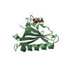
- Assembly
| Deposited unit | 
| |||||||||
|---|---|---|---|---|---|---|---|---|---|---|
| 1 |
| |||||||||
| 2 | 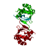
| |||||||||
| Unit cell |
| |||||||||
| Components on special symmetry positions |
|
-Components
| #1: Protein | / HSF 1 / Heat shock transcription factor 1 / HSTF 1 Mass: 13156.973 Da / Num. of mol.: 1 / Fragment: UNP residues 15-120 Source method: isolated from a genetically manipulated source Source: (gene. exp.) Homo sapiens (human) / Gene: HSF1, HSTF1 / Production host:  Escherichia coli (E. coli) / References: UniProt: Q00613 Escherichia coli (E. coli) / References: UniProt: Q00613 |
|---|---|
| #2: Chemical | ChemComp-NA /   Mass: 22.990 Da / Num. of mol.: 1 / Source method: obtained synthetically / Formula: Na Mass: 22.990 Da / Num. of mol.: 1 / Source method: obtained synthetically / Formula: Na |
| #3: Water | ChemComp-HOH / Water Mass: 18.015 Da / Num. of mol.: 149 / Source method: isolated from a natural source / Formula: H2O Mass: 18.015 Da / Num. of mol.: 149 / Source method: isolated from a natural source / Formula: H2O |
-Experimental details
-Experiment
| Experiment | Method: X-RAY DIFFRACTION |
|---|
- Sample preparation
| Crystal | Density Matthews: 2.16 Å3/Da / Density % sol: 43.04 % |
|---|---|
| Crystal grow | Temperature: 293 K / Method: vapor diffusion, hanging drop / pH: 7.2 / Details: 0.2M sodium formate, pH 7.2, 22%(w/v) PEG3350 |
-Data collection
| Diffraction | Mean temperature: 100 K |
|---|---|
| Diffraction source | Source: SYNCHROTRON / Site: SSRF  / Beamline: BL17U / Wavelength: 0.97915 Å / Beamline: BL17U / Wavelength: 0.97915 Å |
| Detector | Type: ADSC QUANTUM 315r / Detector: CCD / Date: Jul 6, 2014 |
| Radiation | Protocol: SINGLE WAVELENGTH / Monochromatic (M) / Laue (L): M / Scattering type: x-ray |
| Radiation wavelength | Wavelength: 0.97915 Å / Relative weight: 1 |
| Reflection | Resolution: 1.7→32.21 Å / Num. obs: 12343 / % possible obs: 99.5 % / Redundancy: 7.1 % / Rmerge(I) obs: 0.12 / Rsym value: 0.12 / Net I/σ(I): 4.1 |
| Reflection shell | Resolution: 1.7→1.73 Å / Redundancy: 7.3 % / Rmerge(I) obs: 0.527 / Mean I/σ(I) obs: 3 / Rsym value: 0.527 / % possible all: 99.8 |
- Processing
| Software |
| ||||||||||||||||||||||||||||||||||||||||||||||||||||||||||||||||||||||
|---|---|---|---|---|---|---|---|---|---|---|---|---|---|---|---|---|---|---|---|---|---|---|---|---|---|---|---|---|---|---|---|---|---|---|---|---|---|---|---|---|---|---|---|---|---|---|---|---|---|---|---|---|---|---|---|---|---|---|---|---|---|---|---|---|---|---|---|---|---|---|---|
| Refinement | Method to determine structure: MOLECULAR REPLACEMENT Starting model: 3HTS Resolution: 1.7→32.21 Å / SU ML: 0.18 / Cross valid method: NONE / σ(F): 1.39 / Phase error: 22.83 / Stereochemistry target values: ML
| ||||||||||||||||||||||||||||||||||||||||||||||||||||||||||||||||||||||
| Solvent computation | Shrinkage radii: 0.9 Å / VDW probe radii: 1.11 Å / Solvent model: FLAT BULK SOLVENT MODEL | ||||||||||||||||||||||||||||||||||||||||||||||||||||||||||||||||||||||
| Refinement step | Cycle: LAST / Resolution: 1.7→32.21 Å
| ||||||||||||||||||||||||||||||||||||||||||||||||||||||||||||||||||||||
| Refine LS restraints |
| ||||||||||||||||||||||||||||||||||||||||||||||||||||||||||||||||||||||
| LS refinement shell |
|
