 Movie
Movie Controller

[English] 日本語
 Yorodumi
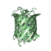
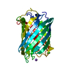
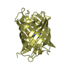
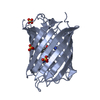
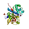
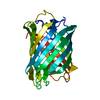
- PDB-5fhv: Crystal structure of mCherry after reaction with 2-mercaptoethanol -
+ Open data

- Basic information
| Entry | Database: PDB / ID: 5fhv | ||||||
|---|---|---|---|---|---|---|---|
| Title | Crystal structure of mCherry after reaction with 2-mercaptoethanol | ||||||
 Components Components | mCherry-BME | ||||||
 Keywords Keywords |  FLUORESCENT PROTEIN / 2-mercaptoethanol / Michael addition FLUORESCENT PROTEIN / 2-mercaptoethanol / Michael addition | ||||||
| Function / homology |  Function and homology informationbioluminescence / generation of precursor metabolites and energy / metal ion binding Function and homology informationbioluminescence / generation of precursor metabolites and energy / metal ion bindingSimilarity search - Function | ||||||
| Biological species |  Discosoma sp. (sea anemone) Discosoma sp. (sea anemone) | ||||||
| Method | X-RAY DIFFRACTION / SYNCHROTRON / MOLECULAR REPLACEMENT / molecular replacement / Resolution: 1.55 Å | ||||||
 Authors Authors | De Zitter, E. / Dedecker, P. / Van Meervelt, L. | ||||||
 Citation Citation | Journal: Proc. Natl. Acad. Sci. U.S.A. / Year: 2017 Title: Efficient switching of mCherry fluorescence using chemical caging. Authors: Cloin, B.M.C. / De Zitter, E. / Salas, D. / Gielen, V. / Folkers, G.E. / Mikhaylova, M. / Bergeler, M. / Krajnik, B. / Harvey, J. / Hoogenraad, C.C. / Van Meervelt, L. / Dedecker, P. / Kapitein, L.C. | ||||||
| History |
|
- Structure visualization
| Structure viewer | Molecule: MolmilJmol/JSmol |
|---|
- Downloads & links
-Download
| PDBx/mmCIF format | 5fhv.cif.gz | 73.9 KB | Display | PDBx/mmCIF format |
|---|---|---|---|---|
| PDB format | pdb5fhv.ent.gz | 51.8 KB | Display | PDB format |
| PDBx/mmJSON format | 5fhv.json.gz | Tree view | PDBx/mmJSON format | |
| Others |  Other downloads Other downloads |
-Validation report
| Arichive directory | https://data.pdbj.org/pub/pdb/validation_reports/fh/5fhvftp://data.pdbj.org/pub/pdb/validation_reports/fh/5fhv | HTTPS FTP |
|---|
-Related structure data
| Related structure data |  2h5qS S: Starting model for refinement |
|---|---|
| Similar structure data |


-Links
 PDBj
PDBj


- Assembly
| Deposited unit | 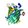
| ||||||||
|---|---|---|---|---|---|---|---|---|---|
| 1 |
| ||||||||
| Unit cell |
|
-Components
| #1: Protein | Mass: 30489.234 Da / Num. of mol.: 1 Source method: isolated from a genetically manipulated source Source: (gene. exp.) Discosoma sp. (sea anemone) / Production host:  Escherichia coli BL21(DE3) (bacteria) / Variant (production host): JM109 / References: UniProt: D0VWW2*PLUS Escherichia coli BL21(DE3) (bacteria) / Variant (production host): JM109 / References: UniProt: D0VWW2*PLUS | ||||
|---|---|---|---|---|---|
| #2: Chemical | ChemComp-PGE / Polyethylene glycol  Mass: 150.173 Da / Num. of mol.: 1 / Source method: obtained synthetically / Formula: C6H14O4 Mass: 150.173 Da / Num. of mol.: 1 / Source method: obtained synthetically / Formula: C6H14O4 | ||||
| #3: Chemical | 2-Mercaptoethanol  Mass: 78.133 Da / Num. of mol.: 3 / Source method: obtained synthetically / Formula: C2H6OS Mass: 78.133 Da / Num. of mol.: 3 / Source method: obtained synthetically / Formula: C2H6OS#4: Chemical | ChemComp-P6G / | Polyethylene glycol  Mass: 282.331 Da / Num. of mol.: 1 / Source method: obtained synthetically / Formula: C12H26O7 / Comment: precipitant*YM Mass: 282.331 Da / Num. of mol.: 1 / Source method: obtained synthetically / Formula: C12H26O7 / Comment: precipitant*YM#5: Water | ChemComp-HOH / | Water Mass: 18.015 Da / Num. of mol.: 315 / Source method: isolated from a natural source / Formula: H2O Mass: 18.015 Da / Num. of mol.: 315 / Source method: isolated from a natural source / Formula: H2O |
-Experimental details
-Experiment
| Experiment | Method: X-RAY DIFFRACTION / Number of used crystals: 1 |
|---|
- Sample preparation
| Crystal | Density Matthews: 2.39 Å3/Da / Density % sol: 48.49 % |
|---|---|
| Crystal grow | Temperature: 289 K / Method: vapor diffusion, sitting drop / pH: 7.4 / Details: 0.2 M MgCl2.6H2O, 0.1 M Tris pH 8.5, 25 % PEG 4000 |
-Data collection
| Diffraction | Mean temperature: 100 K | ||||||||||||||||||
|---|---|---|---|---|---|---|---|---|---|---|---|---|---|---|---|---|---|---|---|
| Diffraction source | Source: SYNCHROTRON / Site: SLS  / Beamline: X06SA / Wavelength: 1 Å / Beamline: X06SA / Wavelength: 1 Å | ||||||||||||||||||
| Detector | Type: DECTRIS PILATUS 2M / Detector: PIXEL / Date: May 23, 2015 | ||||||||||||||||||
| Radiation | Protocol: SINGLE WAVELENGTH / Monochromatic (M) / Laue (L): M / Scattering type: x-ray | ||||||||||||||||||
| Radiation wavelength | Wavelength: 1 Å / Relative weight: 1 | ||||||||||||||||||
| Reflection | Resolution: 1.55→45.381 Å / Num. obs: 34495 / % possible obs: 99.4 % / Redundancy: 3.3 % / Biso Wilson estimate: 13.97 Å2 / CC1/2: 0.999 / Rmerge(I) obs: 0.062 / Net I/σ(I): 14 | ||||||||||||||||||
| Reflection shell |
|
-Phasing
| Phasing | Method: molecular replacement | |||||||||
|---|---|---|---|---|---|---|---|---|---|---|
| Phasing MR |
|
- Processing
| Software |
| |||||||||||||||||||||||||||||||||||||||||||||||||||||||||||||||||||||||||||||||||||||||||||
|---|---|---|---|---|---|---|---|---|---|---|---|---|---|---|---|---|---|---|---|---|---|---|---|---|---|---|---|---|---|---|---|---|---|---|---|---|---|---|---|---|---|---|---|---|---|---|---|---|---|---|---|---|---|---|---|---|---|---|---|---|---|---|---|---|---|---|---|---|---|---|---|---|---|---|---|---|---|---|---|---|---|---|---|---|---|---|---|---|---|---|---|---|
| Refinement | Method to determine structure: MOLECULAR REPLACEMENT Starting model: 2H5Q Resolution: 1.55→45.381 Å / SU ML: 0.17 / Cross valid method: FREE R-VALUE / σ(F): 1.34 / Phase error: 19.14 / Stereochemistry target values: ML
| |||||||||||||||||||||||||||||||||||||||||||||||||||||||||||||||||||||||||||||||||||||||||||
| Solvent computation | Shrinkage radii: 0.9 Å / VDW probe radii: 1.11 Å / Solvent model: FLAT BULK SOLVENT MODEL | |||||||||||||||||||||||||||||||||||||||||||||||||||||||||||||||||||||||||||||||||||||||||||
| Displacement parameters | Biso max: 74.86 Å2 / Biso mean: 17.7512 Å2 / Biso min: 5.7 Å2 | |||||||||||||||||||||||||||||||||||||||||||||||||||||||||||||||||||||||||||||||||||||||||||
| Refinement step | Cycle: final / Resolution: 1.55→45.381 Å
| |||||||||||||||||||||||||||||||||||||||||||||||||||||||||||||||||||||||||||||||||||||||||||
| Refine LS restraints |
| |||||||||||||||||||||||||||||||||||||||||||||||||||||||||||||||||||||||||||||||||||||||||||
| LS refinement shell | Refine-ID: X-RAY DIFFRACTION / Total num. of bins used: 12
|
