 Movie
Movie Controller

[English] 日本語
 Yorodumi
- PDB-2jet: Crystal structure of a trypsin-like mutant (S189D , A226G) chymot... -
+ Open data

- Basic information
| Entry | Database: PDB / ID: 2jet | ||||||
|---|---|---|---|---|---|---|---|
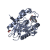
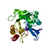
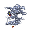
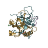
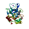
| Title | Crystal structure of a trypsin-like mutant (S189D , A226G) chymotrypsin. | ||||||
 Components Components |
| ||||||
 Keywords Keywords |  HYDROLASE / SUBSTRATE SPECIFICITY / ZYMOGEN / PROTEASE / DIGESTION / SERINE PROTEASE / PROTEIN ENGINEERING HYDROLASE / SUBSTRATE SPECIFICITY / ZYMOGEN / PROTEASE / DIGESTION / SERINE PROTEASE / PROTEIN ENGINEERING | ||||||
| Function / homology |  Function and homology information Function and homology informationActivation of Matrix Metalloproteinases / chymotrypsin / response to food / digestion / serine-type peptidase activity / response to nutrient / response to cytokine / protein catabolic process / response to peptide hormone / response to toxic substance ...Activation of Matrix Metalloproteinases / chymotrypsin / response to food / digestion / serine-type peptidase activity / response to nutrient / response to cytokine / protein catabolic process / response to peptide hormone / response to toxic substance / peptidase activity / positive regulation of apoptotic process / serine-type endopeptidase activity / proteolysis / extracellular regionSimilarity search - Function | ||||||
| Biological species |  RATTUS NORVEGICUS (Norway rat) RATTUS NORVEGICUS (Norway rat) | ||||||
| Method | X-RAY DIFFRACTION / SYNCHROTRON / MOLECULAR REPLACEMENT / Resolution: 2.2 Å | ||||||
 Authors Authors | Jelinek, B. / Katona, G. / Fodor, K. / Venekei, I. / Graf, L. | ||||||
 Citation Citation | Journal: Protein J. / Year: 2008 Title: The Crystal Structure of a Trypsin-Like Mutant Chymotrypsin: The Role of Position 226 in the Activity and Specificity of S189D Chymotrypsin. Authors: Jelinek, B. / Katona, G. / Fodor, K. / Venekei, I. / Graf, L. | ||||||
| History |
| ||||||
| Remark 700 | SHEET DETERMINATION METHOD: DSSP THE SHEETS PRESENTED AS "BA" IN EACH CHAIN ON SHEET RECORDS BELOW ... SHEET DETERMINATION METHOD: DSSP THE SHEETS PRESENTED AS "BA" IN EACH CHAIN ON SHEET RECORDS BELOW IS ACTUALLY AN 8-STRANDED BARREL THIS IS REPRESENTED BY A 9-STRANDED SHEET IN WHICH THE FIRST AND LAST STRANDS ARE IDENTICAL. THE SHEETS PRESENTED AS "BB" IN EACH CHAIN ON SHEET RECORDS BELOW IS ACTUALLY AN 7-STRANDED BARREL THIS IS REPRESENTED BY A 8-STRANDED SHEET IN WHICH THE FIRST AND LAST STRANDS ARE IDENTICAL. |
- Structure visualization
| Structure viewer | Molecule: MolmilJmol/JSmol |
|---|
- Downloads & links
-Download
| PDBx/mmCIF format | 2jet.cif.gz | 57.4 KB | Display | PDBx/mmCIF format |
|---|---|---|---|---|
| PDB format | pdb2jet.ent.gz | 40.5 KB | Display | PDB format |
| PDBx/mmJSON format | 2jet.json.gz | Tree view | PDBx/mmJSON format | |
| Others |  Other downloads Other downloads |
-Validation report
| Arichive directory | https://data.pdbj.org/pub/pdb/validation_reports/je/2jetftp://data.pdbj.org/pub/pdb/validation_reports/je/2jet | HTTPS FTP |
|---|
-Related structure data
| Related structure data | 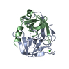 1kdqS S: Starting model for refinement |
|---|---|
| Similar structure data |


-Links
 PDBj
PDBj


- Assembly
| Deposited unit | 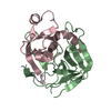
| ||||||||
|---|---|---|---|---|---|---|---|---|---|
| 1 |
| ||||||||
| Unit cell |
|
-Components
| #1: Protein/peptide | Mass: 1544.833 Da / Num. of mol.: 1 / Fragment: RESIDES 19-28 Source method: isolated from a genetically manipulated source Source: (gene. exp.) RATTUS NORVEGICUS (Norway rat) / Organ: PANCREAS / Plasmid: PET17B / Production host:  ESCHERICHIA COLI (E. coli) / Strain (production host): BL21 (DE3) PLYSS / References: UniProt: P07338, chymotrypsin ESCHERICHIA COLI (E. coli) / Strain (production host): BL21 (DE3) PLYSS / References: UniProt: P07338, chymotrypsin | ||
|---|---|---|---|
| #2: Protein | Mass: 13880.524 Da / Num. of mol.: 1 / Fragment: RESIDUES 37-164 Source method: isolated from a genetically manipulated source Source: (gene. exp.) RATTUS NORVEGICUS (Norway rat) / Organ: PANCREAS / Plasmid: PET17B / Production host: ESCHERICHIA COLI (E. coli) / Strain (production host): BL21 (DE3) PLYSS / References: UniProt: P07338, chymotrypsin | ||
| #3: Protein | Mass: 10253.702 Da / Num. of mol.: 1 / Fragment: RESIDUES 165-263 / Mutation: YES Source method: isolated from a genetically manipulated source Source: (gene. exp.) RATTUS NORVEGICUS (Norway rat) / Organ: PANCREAS / Plasmid: PET17B / Production host: ESCHERICHIA COLI (E. coli) / Strain (production host): BL21 (DE3) PLYSS / References: UniProt: P07338, chymotrypsin | ||
| #4: Water | ChemComp-HOH / Water Mass: 18.015 Da / Num. of mol.: 67 / Source method: isolated from a natural source / Formula: H2O Mass: 18.015 Da / Num. of mol.: 67 / Source method: isolated from a natural source / Formula: H2O | ||
| Compound details | ENGINEERED| Sequence details | FIRST FIVE RESIDUE ARE DUE TO THE EXPRESSION | |
-Experimental details
-Experiment
| Experiment | Method: X-RAY DIFFRACTION / Number of used crystals: 1 |
|---|
- Sample preparation
| Crystal | Density Matthews: 1.9 Å3/Da / Density % sol: 35.8 % / Description: NONE |
|---|---|
| Crystal grow | Temperature: 293 K / Method: vapor diffusion / pH: 7 Details: VAPOR DIFFUSION METHOD, 0.1M HEPES, PH 7.0, 30% PEG6000 AND PROTEIN (15 MG/ML) WITH BENZAMIDINE TWOFOLD EXCESS MIXED 1:1, 20 C |
-Data collection
| Diffraction | Mean temperature: 100 K |
|---|---|
| Diffraction source | Source: SYNCHROTRON / Site: ESRF  / Beamline: ID14-2 / Wavelength: 0.933 / Beamline: ID14-2 / Wavelength: 0.933 |
| Detector | Type: ADSC QUANTUM 4 / Detector: CCD / Date: Feb 7, 2005 / Details: MIRRORS |
| Radiation | Monochromator: DIAMOND (111), GE (220) / Protocol: SINGLE WAVELENGTH / Monochromatic (M) / Laue (L): M / Scattering type: x-ray |
| Radiation wavelength | Wavelength: 0.933 Å / Relative weight: 1 |
| Reflection | Resolution: 2.2→64.42 Å / Num. obs: 9753 / % possible obs: 98.5 % / Observed criterion σ(I): 2 / Redundancy: 3.1 % / Rmerge(I) obs: 0.09 / Net I/σ(I): 10.2 |
| Reflection shell | Resolution: 2.2→2.32 Å / Redundancy: 2.8 % / Rmerge(I) obs: 0.44 / Mean I/σ(I) obs: 2.3 / % possible all: 95.8 |
- Processing
| Software |
| ||||||||||||||||||||||||||||||||||||||||||||||||||||||||||||||||||||||||||||||||||||||||||||||||||||||||||||||||||||||||||||||||||||||||||||||||||||||||||||||||||||||||||||||||||||||
|---|---|---|---|---|---|---|---|---|---|---|---|---|---|---|---|---|---|---|---|---|---|---|---|---|---|---|---|---|---|---|---|---|---|---|---|---|---|---|---|---|---|---|---|---|---|---|---|---|---|---|---|---|---|---|---|---|---|---|---|---|---|---|---|---|---|---|---|---|---|---|---|---|---|---|---|---|---|---|---|---|---|---|---|---|---|---|---|---|---|---|---|---|---|---|---|---|---|---|---|---|---|---|---|---|---|---|---|---|---|---|---|---|---|---|---|---|---|---|---|---|---|---|---|---|---|---|---|---|---|---|---|---|---|---|---|---|---|---|---|---|---|---|---|---|---|---|---|---|---|---|---|---|---|---|---|---|---|---|---|---|---|---|---|---|---|---|---|---|---|---|---|---|---|---|---|---|---|---|---|---|---|---|---|
| Refinement | Method to determine structure: MOLECULAR REPLACEMENT Starting model: PDB ENTRY 1KDQ Resolution: 2.2→43.23 Å / Cor.coef. Fo:Fc: 0.895 / Cor.coef. Fo:Fc free: 0.834 / SU B: 24.796 / SU ML: 0.332 / TLS residual ADP flag: LIKELY RESIDUAL / Cross valid method: THROUGHOUT / ESU R: 0.558 / ESU R Free: 0.328 / Stereochemistry target values: MAXIMUM LIKELIHOOD Details: HYDROGENS HAVE BEEN ADDED IN THE RIDING POSITIONS. DISORDERED SIDECHAINS WITH 0 OCCUPANCY
| ||||||||||||||||||||||||||||||||||||||||||||||||||||||||||||||||||||||||||||||||||||||||||||||||||||||||||||||||||||||||||||||||||||||||||||||||||||||||||||||||||||||||||||||||||||||
| Solvent computation | Ion probe radii: 0.8 Å / Shrinkage radii: 0.8 Å / VDW probe radii: 1.2 Å / Solvent model: BABINET MODEL WITH MASK | ||||||||||||||||||||||||||||||||||||||||||||||||||||||||||||||||||||||||||||||||||||||||||||||||||||||||||||||||||||||||||||||||||||||||||||||||||||||||||||||||||||||||||||||||||||||
| Displacement parameters | Biso mean: 44.1 Å2
| ||||||||||||||||||||||||||||||||||||||||||||||||||||||||||||||||||||||||||||||||||||||||||||||||||||||||||||||||||||||||||||||||||||||||||||||||||||||||||||||||||||||||||||||||||||||
| Refinement step | Cycle: LAST / Resolution: 2.2→43.23 Å
| ||||||||||||||||||||||||||||||||||||||||||||||||||||||||||||||||||||||||||||||||||||||||||||||||||||||||||||||||||||||||||||||||||||||||||||||||||||||||||||||||||||||||||||||||||||||
| Refine LS restraints |
|