 Movie
Movie Controller


+ Open data

- Basic information
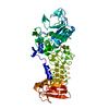
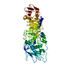
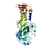
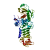
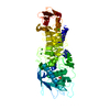
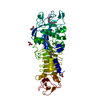
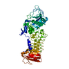
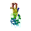
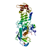
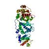
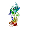
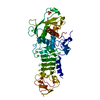
| Entry | Database: PDB / ID: 1srp | ||||||
|---|---|---|---|---|---|---|---|
| Title | STRUCTURAL ANALYSIS OF SERRATIA PROTEASE | ||||||
 Components Components | SERRALYSIN | ||||||
 Keywords Keywords | HYDROLASE (METALLOPROTEASE) | ||||||
| Function / homology |  Function and homology informationserralysin / extracellular matrix / metalloendopeptidase activity / calcium ion binding / proteolysis / extracellular space / zinc ion binding Function and homology informationserralysin / extracellular matrix / metalloendopeptidase activity / calcium ion binding / proteolysis / extracellular space / zinc ion bindingSimilarity search - Function | ||||||
| Biological species |  Serratia sp. (bacteria) Serratia sp. (bacteria) | ||||||
| Method | X-RAY DIFFRACTION / Resolution: 2 Å | ||||||
 Authors Authors | Hamada, K. / Hiramatsu, H. / Katsuya, Y. / Hata, Y. / Katsube, Y. | ||||||
 Citation Citation | Journal: J.Biochem.(Tokyo) / Year: 1996 Title: Crystal structure of Serratia protease, a zinc-dependent proteinase from Serratia sp. E-15, containing a beta-sheet coil motif at 2.0 A resolution. Authors: Hamada, K. / Hata, Y. / Katsuya, Y. / Hiramatsu, H. / Fujiwara, T. / Katsube, Y. #1: Journal: Photon Factory Activity Report / Year: 1992Title: Structural Studies of Serratia Protease by X-Ray Analysis Authors: Hamada, K. / Hiramatsu, H. / Fujiwara, T. / Katsuya, Y. / Hata, Y. / Katsube, Y. #2: Journal: J.Biochem.(Tokyo) / Year: 1985Title: Preliminary X-Ray Studies on Serratia Protease Authors: Katsuya, Y. / Hamada, K. / Hata, Y. / Tanaka, N. / Sato, M. / Katsube, Y. / Katiuchi, K. / Miyata, K. | ||||||
| History |
|
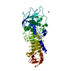
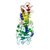
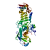
- Structure visualization
| Structure viewer | Molecule: MolmilJmol/JSmol |
|---|
- Downloads & links
-Download
| PDBx/mmCIF format | 1srp.cif.gz | 105.1 KB | Display | PDBx/mmCIF format |
|---|---|---|---|---|
| PDB format | pdb1srp.ent.gz | 80.3 KB | Display | PDB format |
| PDBx/mmJSON format | 1srp.json.gz | Tree view | PDBx/mmJSON format | |
| Others |  Other downloads Other downloads |
-Validation report
| Arichive directory | https://data.pdbj.org/pub/pdb/validation_reports/sr/1srpftp://data.pdbj.org/pub/pdb/validation_reports/sr/1srp | HTTPS FTP |
|---|
-Related structure data
| Similar structure data |
|---|


-Links
 PDBj
PDBj


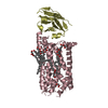
- Assembly
| Deposited unit | 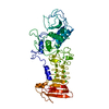
| ||||||||
|---|---|---|---|---|---|---|---|---|---|
| 1 |
| ||||||||
| Unit cell |
| ||||||||
| Atom site foot note | 1: SEVEN CALCIUM IONS, RESIDUES CA 911 - CA 917, ARE LIGATED TO THE PROTEIN. |
-Components
| #1: Protein | Mass: 50493.312 Da / Num. of mol.: 1 Source method: isolated from a genetically manipulated source Source: (gene. exp.) Serratia sp. (bacteria) / Strain: E-15 / References: UniProt: P07268, serralysin | ||||
|---|---|---|---|---|---|
| #2: Chemical | ChemComp-CA /   Mass: 40.078 Da / Num. of mol.: 7 / Source method: obtained synthetically / Formula: Ca Mass: 40.078 Da / Num. of mol.: 7 / Source method: obtained synthetically / Formula: Ca#3: Chemical | ChemComp-ZN / |   Mass: 65.409 Da / Num. of mol.: 1 / Source method: obtained synthetically / Formula: Zn Mass: 65.409 Da / Num. of mol.: 1 / Source method: obtained synthetically / Formula: Zn#4: Water | ChemComp-HOH / | Water Mass: 18.015 Da / Num. of mol.: 212 / Source method: isolated from a natural source / Formula: H2O Mass: 18.015 Da / Num. of mol.: 212 / Source method: isolated from a natural source / Formula: H2O |
-Experimental details
-Experiment
| Experiment | Method: X-RAY DIFFRACTION |
|---|
- Sample preparation
| Crystal | Density Matthews: 3.48 Å3/Da / Density % sol: 64.61 % | ||||||||||||||||||||
|---|---|---|---|---|---|---|---|---|---|---|---|---|---|---|---|---|---|---|---|---|---|
| Crystal | *PLUS Density % sol: 67 % | ||||||||||||||||||||
| Crystal grow | *PLUS Method: microdialysis | ||||||||||||||||||||
| Components of the solutions | *PLUS
|
-Data collection
| Reflection | Num. obs: 42930 / % possible obs: 88.2 % / Observed criterion σ(I): 0 |
|---|
- Processing
| Software | Name: GPRLSA / Classification: refinement | ||||||||||||||||||||||||||||||||||||||||||||||||||||||||||||||||||||||||||||||||||||
|---|---|---|---|---|---|---|---|---|---|---|---|---|---|---|---|---|---|---|---|---|---|---|---|---|---|---|---|---|---|---|---|---|---|---|---|---|---|---|---|---|---|---|---|---|---|---|---|---|---|---|---|---|---|---|---|---|---|---|---|---|---|---|---|---|---|---|---|---|---|---|---|---|---|---|---|---|---|---|---|---|---|---|---|---|---|
| Refinement | Resolution: 2→8 Å / σ(F): 2
| ||||||||||||||||||||||||||||||||||||||||||||||||||||||||||||||||||||||||||||||||||||
| Displacement parameters | Biso mean: 18.97 Å2 | ||||||||||||||||||||||||||||||||||||||||||||||||||||||||||||||||||||||||||||||||||||
| Refinement step | Cycle: LAST / Resolution: 2→8 Å
| ||||||||||||||||||||||||||||||||||||||||||||||||||||||||||||||||||||||||||||||||||||
| Refine LS restraints |
| ||||||||||||||||||||||||||||||||||||||||||||||||||||||||||||||||||||||||||||||||||||
| Refinement | *PLUS Num. reflection obs: 38044 / σ(F): 3 / Rfactor obs: 0.184 | ||||||||||||||||||||||||||||||||||||||||||||||||||||||||||||||||||||||||||||||||||||
| Solvent computation | *PLUS | ||||||||||||||||||||||||||||||||||||||||||||||||||||||||||||||||||||||||||||||||||||
| Displacement parameters | *PLUS |
