 Movie
Movie Controller

[English] 日本語
 Yorodumi
- PDB-1qgh: THE X-RAY STRUCTURE OF THE UNUSUAL DODECAMERIC FERRITIN FROM LIST... -
+ Open data

- Basic information
| Entry | Database: PDB / ID: 1qgh | ||||||
|---|---|---|---|---|---|---|---|
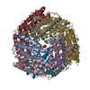
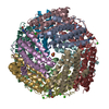
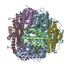
| Title | THE X-RAY STRUCTURE OF THE UNUSUAL DODECAMERIC FERRITIN FROM LISTERIA INNOCUA, REVEALS A NOVEL INTERSUBUNIT IRON BINDING SITE. | ||||||
 Components Components | NON-HEME IRON-CONTAINING FERRITIN | ||||||
 Keywords Keywords | METAL TRANSPORT /  FERRITIN FERRITIN | ||||||
| Function / homology |  Function and homology informationOxidoreductases; Oxidizing metal ions / oxidoreductase activity, acting on metal ions / ferric iron binding / intracellular iron ion homeostasis / cytoplasm Function and homology informationOxidoreductases; Oxidizing metal ions / oxidoreductase activity, acting on metal ions / ferric iron binding / intracellular iron ion homeostasis / cytoplasmSimilarity search - Function | ||||||
| Biological species |  Listeria innocua (bacteria) Listeria innocua (bacteria) | ||||||
| Method | X-RAY DIFFRACTION / SYNCHROTRON / MOLECULAR REPLACEMENT / Resolution: 2.35 Å | ||||||
 Authors Authors | Ilari, A. / Stefanini, S. / Chiancone, E. / Tsernoglou, D. | ||||||
 Citation Citation | Journal: Nat.Struct.Biol. / Year: 2000 Title: The dodecameric ferritin from Listeria innocua contains a novel intersubunit iron-binding site. Authors: Ilari, A. / Stefanini, S. / Chiancone, E. / Tsernoglou, D. | ||||||
| History |
|
- Structure visualization
| Structure viewer | Molecule: MolmilJmol/JSmol |
|---|
- Downloads & links
-Download
| PDBx/mmCIF format | 1qgh.cif.gz | 371.8 KB | Display | PDBx/mmCIF format |
|---|---|---|---|---|
| PDB format | pdb1qgh.ent.gz | 302.7 KB | Display | PDB format |
| PDBx/mmJSON format | 1qgh.json.gz | Tree view | PDBx/mmJSON format | |
| Others |  Other downloads Other downloads |
-Validation report
| Arichive directory | https://data.pdbj.org/pub/pdb/validation_reports/qg/1qghftp://data.pdbj.org/pub/pdb/validation_reports/qg/1qgh | HTTPS FTP |
|---|
-Related structure data
| Related structure data | 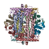 1dpsS S: Starting model for refinement |
|---|---|
| Similar structure data |


-Links
 PDBj
PDBj


- Assembly
| Deposited unit | 
| ||||||||||||||||||||||||||||||||||||||||||||||||
|---|---|---|---|---|---|---|---|---|---|---|---|---|---|---|---|---|---|---|---|---|---|---|---|---|---|---|---|---|---|---|---|---|---|---|---|---|---|---|---|---|---|---|---|---|---|---|---|---|---|
| 1 |
| ||||||||||||||||||||||||||||||||||||||||||||||||
| Unit cell |
| ||||||||||||||||||||||||||||||||||||||||||||||||
| Noncrystallographic symmetry (NCS) | NCS oper:
|
-Components
| #1: Protein | Mass: 18070.553 Da / Num. of mol.: 12 / Source method: isolated from a natural source / Source: (natural) Listeria innocua (bacteria) / References: UniProt: P80725#2: Chemical | ChemComp-FE / Iron  Mass: 55.845 Da / Num. of mol.: 12 / Source method: obtained synthetically / Formula: Fe Mass: 55.845 Da / Num. of mol.: 12 / Source method: obtained synthetically / Formula: Fe#3: Water | ChemComp-HOH / | Water Mass: 18.015 Da / Num. of mol.: 479 / Source method: isolated from a natural source / Formula: H2O Mass: 18.015 Da / Num. of mol.: 479 / Source method: isolated from a natural source / Formula: H2O |
|---|
-Experimental details
-Experiment
| Experiment | Method: X-RAY DIFFRACTION / Number of used crystals: 1 |
|---|
- Sample preparation
| Crystal | Density Matthews: 2.41 Å3/Da / Density % sol: 48.86 % | ||||||||||||||||||||
|---|---|---|---|---|---|---|---|---|---|---|---|---|---|---|---|---|---|---|---|---|---|
| Crystal grow | pH: 5.8 Details: PEG 1000 AT 19-25 %, MES 0.1 M AT PH 5.0-6.2, pH 5.8 | ||||||||||||||||||||
| Crystal grow | *PLUS Temperature: 293 K / Method: vapor diffusion, sitting drop | ||||||||||||||||||||
| Components of the solutions | *PLUS
|
-Data collection
| Diffraction | Mean temperature: 100 K |
|---|---|
| Diffraction source | Source: SYNCHROTRON / Site: EMBL/DESY, HAMBURG  / Beamline: BW7B / Wavelength: 0.8373 / Beamline: BW7B / Wavelength: 0.8373 |
| Detector | Date: Dec 1, 1997 |
| Radiation | Protocol: SINGLE WAVELENGTH / Monochromatic (M) / Laue (L): M / Scattering type: x-ray |
| Radiation wavelength | Wavelength: 0.8373 Å / Relative weight: 1 |
| Reflection | Resolution: 2.35→30 Å / Num. obs: 80150 / % possible obs: 91.3 % / Redundancy: 3.8 % / Rsym value: 0.077 |
| Reflection | *PLUS Rmerge(I) obs: 0.077 |
- Processing
| Software |
| ||||||||||||||||||||||||||||||
|---|---|---|---|---|---|---|---|---|---|---|---|---|---|---|---|---|---|---|---|---|---|---|---|---|---|---|---|---|---|---|---|
| Refinement | Method to determine structure: MOLECULAR REPLACEMENT Starting model: 1DPS Resolution: 2.35→10 Å / σ(F): 0
| ||||||||||||||||||||||||||||||
| Refinement step | Cycle: LAST / Resolution: 2.35→10 Å
| ||||||||||||||||||||||||||||||
| Refine LS restraints |
| ||||||||||||||||||||||||||||||
| Software | *PLUS Name: TNT / Version: 5E / Classification: refinement | ||||||||||||||||||||||||||||||
| Refine LS restraints | *PLUS
|
