 Movie
Movie Controller


+ Open data

- Basic information
| Entry | Database: PDB / ID: 1g2t | ||||||
|---|---|---|---|---|---|---|---|
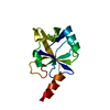
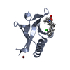
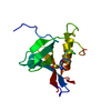
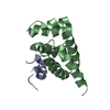
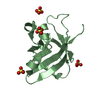
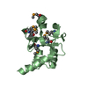
| Title | SOLUTION STRUCTURE OF EOTAXIN-3 | ||||||
 Components Components | EOTAXIN-3 | ||||||
 Keywords Keywords | CYTOKINE / beta-beta-beta-alpha helix | ||||||
| Function / homology |  Function and homology information Function and homology informationCCR3 chemokine receptor binding / CX3C chemokine receptor binding / CCR chemokine receptor binding / lymphocyte chemotaxis / positive regulation of chemotaxis / T cell chemotaxis / eosinophil chemotaxis / chemokine-mediated signaling pathway / chemokine activity / positive regulation of actin filament polymerization ...CCR3 chemokine receptor binding / CX3C chemokine receptor binding / CCR chemokine receptor binding / lymphocyte chemotaxis / positive regulation of chemotaxis / T cell chemotaxis / eosinophil chemotaxis / chemokine-mediated signaling pathway / chemokine activity / positive regulation of actin filament polymerization / monocyte chemotaxis / cellular response to interleukin-1 / positive regulation of endothelial cell proliferation / neutrophil chemotaxis / positive regulation of GTPase activity / cellular response to type II interferon / chemotaxis / cell-cell signaling / cellular response to tumor necrosis factor / receptor ligand activity / positive regulation of ERK1 and ERK2 cascade / positive regulation of cell migration / inflammatory response / G protein-coupled receptor signaling pathway / signal transduction / extracellular spaceSimilarity search - Function | ||||||
| Biological species |  Homo sapiens (human) Homo sapiens (human) | ||||||
| Method | SOLUTION NMR / distance geometry-simulated annealing protocol implemented in XPLOR | ||||||
| Model type details | minimized average | ||||||
 Authors Authors | Ye, J. / Mayer, K.L. / Mayer, M.R. / Stone, M.J. | ||||||
 Citation Citation | Journal: Biochemistry / Year: 2001 Title: NMR solution structure and backbone dynamics of the CC chemokine eotaxin-3. Authors: Ye, J. / Mayer, K.L. / Mayer, M.R. / Stone, M.J. | ||||||
| History |
|
- Structure visualization
| Structure viewer | Molecule: MolmilJmol/JSmol |
|---|
- Downloads & links
-Download
| PDBx/mmCIF format | 1g2t.cif.gz | 463 KB | Display | PDBx/mmCIF format |
|---|---|---|---|---|
| PDB format | pdb1g2t.ent.gz | 402.6 KB | Display | PDB format |
| PDBx/mmJSON format | 1g2t.json.gz | Tree view | PDBx/mmJSON format | |
| Others |  Other downloads Other downloads |
-Validation report
| Arichive directory | https://data.pdbj.org/pub/pdb/validation_reports/g2/1g2tftp://data.pdbj.org/pub/pdb/validation_reports/g2/1g2t | HTTPS FTP |
|---|
-Related structure data


-Links
 PDBj
PDBj
- Assembly
| Deposited unit | 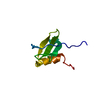
| |||||||||
|---|---|---|---|---|---|---|---|---|---|---|
| 1 |
| |||||||||
| NMR ensembles |
|
-Components
| #1: Protein | / SMALL INDUCIBLE CYTOKINE A26 / MACROPHAGE INFLAMMATORY PROTEIN 4-ALPHA Mass: 8412.857 Da / Num. of mol.: 1 Source method: isolated from a genetically manipulated source Source: (gene. exp.) Homo sapiens (human) / Gene: EOTAXIN-3 / Organ: LUNG / Plasmid: PET28A+ / Species (production host): Escherichia coli / Production host:  Escherichia coli BL21(DE3) (bacteria) / Strain (production host): BL21(DE3) / References: UniProt: Q9Y258 Escherichia coli BL21(DE3) (bacteria) / Strain (production host): BL21(DE3) / References: UniProt: Q9Y258 |
|---|
-Experimental details
-Experiment
| Experiment | Method: SOLUTION NMR | ||||||||||||||||||||||||
|---|---|---|---|---|---|---|---|---|---|---|---|---|---|---|---|---|---|---|---|---|---|---|---|---|---|
| NMR experiment |
|
- Sample preparation
| Details |
| |||||||||||||||
|---|---|---|---|---|---|---|---|---|---|---|---|---|---|---|---|---|
| Sample conditions | Ionic strength: 20 mM / pH: 5 / Pressure: ambient / Temperature: 303 K | |||||||||||||||
| Crystal grow | *PLUS Method: other / Details: NMR |
-NMR measurement
| Radiation | Protocol: SINGLE WAVELENGTH / Monochromatic (M) / Laue (L): M |
|---|---|
| Radiation wavelength | Relative weight: 1 |
| NMR spectrometer | Type: Varian UNITY / Manufacturer: Varian / Model: UNITY / Field strength: 500 MHz |
- Processing
| NMR software |
| ||||||||||||||||||||||||
|---|---|---|---|---|---|---|---|---|---|---|---|---|---|---|---|---|---|---|---|---|---|---|---|---|---|
| Refinement | Method: distance geometry-simulated annealing protocol implemented in XPLOR Software ordinal: 1 Details: The structures are based on a total of 1250 restraints, 1157 are NOE-derived distance constraints, 69 dihedral angle restraints, 17 distance restraints from hydrogn bonds. | ||||||||||||||||||||||||
| NMR representative | Selection criteria: minimized average structure | ||||||||||||||||||||||||
| NMR ensemble | Conformer selection criteria: structures with the lowest energy Conformers calculated total number: 100 / Conformers submitted total number: 20 |
