 Movie
Movie Controller

[English] 日本語
 Yorodumi
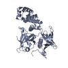
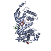
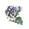
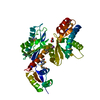
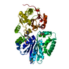
- PDB-1buh: CRYSTAL STRUCTURE OF THE HUMAN CDK2 KINASE COMPLEX WITH CELL CYCL... -
+ Open data

- Basic information
| Entry | Database: PDB / ID: 1buh | ||||||
|---|---|---|---|---|---|---|---|
| Title | CRYSTAL STRUCTURE OF THE HUMAN CDK2 KINASE COMPLEX WITH CELL CYCLE-REGULATORY PROTEIN CKSHS1 | ||||||
 Components Components |
| ||||||
 Keywords Keywords |  TRANSFERASE TRANSFERASE | ||||||
| Function / homology |  Function and homology information Function and homology informationmitotic cell cycle phase transition / cyclin-dependent protein serine/threonine kinase activator activity / SCF ubiquitin ligase complex / cyclin A1-CDK2 complex / cyclin E2-CDK2 complex / cyclin E1-CDK2 complex / cyclin A2-CDK2 complex / positive regulation of DNA-templated DNA replication initiation / G2 Phase / cyclin-dependent protein kinase activity ...mitotic cell cycle phase transition / cyclin-dependent protein serine/threonine kinase activator activity / SCF ubiquitin ligase complex / cyclin A1-CDK2 complex / cyclin E2-CDK2 complex / cyclin E1-CDK2 complex / cyclin A2-CDK2 complex / positive regulation of DNA-templated DNA replication initiation / G2 Phase / cyclin-dependent protein kinase activity / Y chromosome / Phosphorylation of proteins involved in G1/S transition by active Cyclin E:Cdk2 complexes / positive regulation of heterochromatin formation / p53-Dependent G1 DNA Damage Response / X chromosome / PTK6 Regulates Cell Cycle / regulation of mitotic cell cycle / regulation of anaphase-promoting complex-dependent catabolic process / Defective binding of RB1 mutants to E2F1,(E2F2, E2F3) / centriole replication / Regulation of APC/C activators between G1/S and early anaphase / centrosome duplication / Telomere Extension By Telomerase / G0 and Early G1 / Activation of the pre-replicative complex / cyclin-dependent protein kinase holoenzyme complex / cellular response to nitric oxide / Cajal body / cyclin-dependent kinase / cyclin-dependent protein serine/threonine kinase activity / TP53 Regulates Transcription of Genes Involved in G1 Cell Cycle Arrest / Activation of ATR in response to replication stress / Cyclin E associated events during G1/S transition / Cyclin A/B1/B2 associated events during G2/M transition / Cyclin A:Cdk2-associated events at S phase entry / condensed chromosome / mitotic G1 DNA damage checkpoint signaling / regulation of G2/M transition of mitotic cell cycle / cyclin binding / post-translational protein modification / meiotic cell cycle / ubiquitin binding / male germ cell nucleus / response to organic substance / G1/S transition of mitotic cell cycle / potassium ion transport / DNA Damage/Telomere Stress Induced Senescence / CDK-mediated phosphorylation and removal of Cdc6 / SCF(Skp2)-mediated degradation of p27/p21 / Meiotic recombination / Orc1 removal from chromatin / Transcriptional regulation of granulopoiesis / Cyclin D associated events in G1 / G2/M transition of mitotic cell cycle / cellular senescence / Regulation of TP53 Degradation / nuclear envelope / Factors involved in megakaryocyte development and platelet production / Processing of DNA double-strand break ends / histone binding / fibroblast proliferation / Senescence-Associated Secretory Phenotype (SASP) / regulation of gene expression / peptidyl-serine phosphorylation / Ras protein signal transduction / Regulation of TP53 Activity through Phosphorylation / transcription regulator complex / DNA replication / chromosome, telomeric region / endosome / chromatin remodeling / cell division / protein domain specific binding / protein phosphorylation / DNA repair / protein serine kinase activity / centrosome / protein serine/threonine kinase activity / DNA-templated transcription / positive regulation of cell population proliferation / regulation of DNA-templated transcription / protein kinase binding / positive regulation of DNA-templated transcription / negative regulation of transcription by RNA polymerase II / magnesium ion binding / signal transduction / nucleoplasm / ATP binding / nucleus / cytosol / cytoplasmSimilarity search - Function | ||||||
| Biological species |  Homo sapiens (human) Homo sapiens (human) | ||||||
| Method | X-RAY DIFFRACTION / MOLECULAR REPLACEMENT / Resolution: 2.6 Å | ||||||
 Authors Authors | Bourne, Y. / Tainer, J.A. | ||||||
 Citation Citation | Journal: Cell(Cambridge,Mass.) / Year: 1996 Title: Crystal structure and mutational analysis of the human CDK2 kinase complex with cell cycle-regulatory protein CksHs1. Authors: Bourne, Y. / Watson, M.H. / Hickey, M.J. / Holmes, W. / Rocque, W. / Reed, S.I. / Tainer, J.A. | ||||||
| History |
|
- Structure visualization
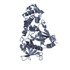
| Structure viewer | Molecule: MolmilJmol/JSmol |
|---|
- Downloads & links
-Download
| PDBx/mmCIF format | 1buh.cif.gz | 85.9 KB | Display | PDBx/mmCIF format |
|---|---|---|---|---|
| PDB format | pdb1buh.ent.gz | 64.8 KB | Display | PDB format |
| PDBx/mmJSON format | 1buh.json.gz | Tree view | PDBx/mmJSON format | |
| Others |  Other downloads Other downloads |
-Validation report
| Arichive directory | https://data.pdbj.org/pub/pdb/validation_reports/bu/1buhftp://data.pdbj.org/pub/pdb/validation_reports/bu/1buh | HTTPS FTP |
|---|
-Related structure data
| Similar structure data |
|---|


-Links
 PDBj
PDBj


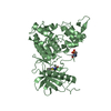
- Assembly
| Deposited unit | 
| ||||||||
|---|---|---|---|---|---|---|---|---|---|
| 1 |
| ||||||||
| Unit cell |
|
-Components
| #1: Protein | Mass: 33976.488 Da / Num. of mol.: 1 Source method: isolated from a genetically manipulated source Source: (gene. exp.) Homo sapiens (human) / Production host:  Trichoplusia ni (cabbage looper) / References: UniProt: P24941 Trichoplusia ni (cabbage looper) / References: UniProt: P24941 |
|---|---|
| #2: Protein | Mass: 9679.211 Da / Num. of mol.: 1 Source method: isolated from a genetically manipulated source Source: (gene. exp.) Homo sapiens (human) / Plasmid: pRK171 / Production host:  Escherichia coli (E. coli) / References: UniProt: P61024 Escherichia coli (E. coli) / References: UniProt: P61024 |
| #3: Water | ChemComp-HOH / Water Mass: 18.015 Da / Num. of mol.: 127 / Source method: isolated from a natural source / Formula: H2O Mass: 18.015 Da / Num. of mol.: 127 / Source method: isolated from a natural source / Formula: H2O |
-Experimental details
-Experiment
| Experiment | Method: X-RAY DIFFRACTION / Number of used crystals: 1 |
|---|
- Sample preparation
| Crystal | Density Matthews: 2.5 Å3/Da / Density % sol: 50.7 % | ||||||||||||||||||||||||||||||
|---|---|---|---|---|---|---|---|---|---|---|---|---|---|---|---|---|---|---|---|---|---|---|---|---|---|---|---|---|---|---|---|
| Crystal grow | pH: 7.5 Details: 17-19% MEPEG 5K, 100 MM TRIS-HCL PH 7.5, 100 MM KCL | ||||||||||||||||||||||||||||||
| Crystal | *PLUS | ||||||||||||||||||||||||||||||
| Crystal grow | *PLUS Temperature: 20 ℃ / Method: vapor diffusion | ||||||||||||||||||||||||||||||
| Components of the solutions | *PLUS
|
-Data collection
| Diffraction | Mean temperature: 100 K |
|---|---|
| Diffraction source | Source: ROTATING ANODE / Type: SIEMENS / Wavelength: 1.5418 |
| Detector | Type: MARRESEARCH / Detector: IMAGE PLATE / Date: Dec 15, 1995 |
| Radiation | Monochromator: NI FILTER / Protocol: SINGLE WAVELENGTH / Monochromatic (M) / Laue (L): M / Scattering type: x-ray |
| Radiation wavelength | Wavelength: 1.5418 Å / Relative weight: 1 |
| Reflection | Resolution: 2.6→20 Å / Num. obs: 84909 / % possible obs: 97 % / Rmerge(I) obs: 0.1 / Rsym value: 0.1 |
| Reflection | *PLUS Num. obs: 13023 / Num. measured all: 84909 / Rmerge(I) obs: 0.1 |
- Processing
| Software |
| ||||||||||||||||||||||||||||||||||||||||||||||||||||||||||||
|---|---|---|---|---|---|---|---|---|---|---|---|---|---|---|---|---|---|---|---|---|---|---|---|---|---|---|---|---|---|---|---|---|---|---|---|---|---|---|---|---|---|---|---|---|---|---|---|---|---|---|---|---|---|---|---|---|---|---|---|---|---|
| Refinement | Method to determine structure: MOLECULAR REPLACEMENT Starting model: PERSONAL COMMUNICATION Resolution: 2.6→20 Å / Cross valid method: THROUGHOUT / σ(F): 0
| ||||||||||||||||||||||||||||||||||||||||||||||||||||||||||||
| Displacement parameters | Biso mean: 24 Å2 | ||||||||||||||||||||||||||||||||||||||||||||||||||||||||||||
| Refinement step | Cycle: LAST / Resolution: 2.6→20 Å
| ||||||||||||||||||||||||||||||||||||||||||||||||||||||||||||
| Refine LS restraints |
| ||||||||||||||||||||||||||||||||||||||||||||||||||||||||||||
| LS refinement shell | Resolution: 2.6→2.7 Å / Total num. of bins used: 8
| ||||||||||||||||||||||||||||||||||||||||||||||||||||||||||||
| Xplor file | Serial no: 1 / Param file: PARHCSDX.PRO / Topol file: TOPHCSDX.PRO | ||||||||||||||||||||||||||||||||||||||||||||||||||||||||||||
| Software | *PLUS Name: X-PLOR / Version: 3.8 / Classification: refinement | ||||||||||||||||||||||||||||||||||||||||||||||||||||||||||||
| Refinement | *PLUS Highest resolution: 2.6 Å / Lowest resolution: 20 Å / σ(F): 0 / % reflection Rfree: 5 % / Rfactor obs: 0.19 / Rfactor Rfree: 0.25 / Rfactor Rwork: 0.19 | ||||||||||||||||||||||||||||||||||||||||||||||||||||||||||||
| Solvent computation | *PLUS | ||||||||||||||||||||||||||||||||||||||||||||||||||||||||||||
| Displacement parameters | *PLUS Biso mean: 24 Å2 | ||||||||||||||||||||||||||||||||||||||||||||||||||||||||||||
| Refine LS restraints | *PLUS
| ||||||||||||||||||||||||||||||||||||||||||||||||||||||||||||
| LS refinement shell | *PLUS Highest resolution: 2.6 Å / Lowest resolution: 2.7 Å / Rfactor Rfree: 0.31 / % reflection Rfree: 5 % / Rfactor Rwork: 0.28 |
