 Movie
Movie Controller


+ Open data

- Basic information
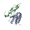
| Entry | Database: PDB / ID: 1a0a | ||||||
|---|---|---|---|---|---|---|---|
| Title | PHOSPHATE SYSTEM POSITIVE REGULATORY PROTEIN PHO4/DNA COMPLEX | ||||||
 Components Components |
| ||||||
 Keywords Keywords | TRANSCRIPTION/DNA /  TRANSCRIPTION FACTOR / BASIC HELIX LOOP HELIX / COMPLEX (TRANSCRIPTION FACTOR-DNA) / TRANSCRIPTION-DNA COMPLEX TRANSCRIPTION FACTOR / BASIC HELIX LOOP HELIX / COMPLEX (TRANSCRIPTION FACTOR-DNA) / TRANSCRIPTION-DNA COMPLEX | ||||||
| Function / homology |  Function and homology information Function and homology informationpositive regulation of phosphate metabolic process / cellular response to phosphate starvation / sequence-specific DNA binding / protein dimerization activity / chromatin remodeling / DNA-binding transcription factor activity / positive regulation of transcription by RNA polymerase II / nucleus / cytoplasmSimilarity search - Function | ||||||
| Biological species |  Saccharomyces cerevisiae (brewer's yeast) Saccharomyces cerevisiae (brewer's yeast) | ||||||
| Method | X-RAY DIFFRACTION / SYNCHROTRON / MIR / Resolution: 2.8 Å | ||||||
 Authors Authors | Shimizu, T. / Toumoto, A. / Ihara, K. / Shimizu, M. / Kyogoku, Y. / Ogawa, N. / Oshima, Y. / Hakoshima, T. | ||||||
 Citation Citation | Journal: EMBO J. / Year: 1997 Title: Crystal structure of PHO4 bHLH domain-DNA complex: flanking base recognition. Authors: Shimizu, T. / Toumoto, A. / Ihara, K. / Shimizu, M. / Kyogoku, Y. / Ogawa, N. / Oshima, Y. / Hakoshima, T. | ||||||
| History |
|
- Structure visualization
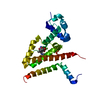
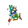
| Structure viewer | Molecule: MolmilJmol/JSmol |
|---|
- Downloads & links
-Download
| PDBx/mmCIF format | 1a0a.cif.gz | 58.2 KB | Display | PDBx/mmCIF format |
|---|---|---|---|---|
| PDB format | pdb1a0a.ent.gz | 40.1 KB | Display | PDB format |
| PDBx/mmJSON format | 1a0a.json.gz | Tree view | PDBx/mmJSON format | |
| Others |  Other downloads Other downloads |
-Validation report
| Arichive directory | https://data.pdbj.org/pub/pdb/validation_reports/a0/1a0aftp://data.pdbj.org/pub/pdb/validation_reports/a0/1a0a | HTTPS FTP |
|---|
-Related structure data
| Similar structure data |
|---|


-Links
 PDBj
PDBj


- Assembly
| Deposited unit | 
| ||||||||
|---|---|---|---|---|---|---|---|---|---|
| 1 |
| ||||||||
| Unit cell |
|
-Components
| #1: DNA chain | Mass: 5212.386 Da / Num. of mol.: 1 / Fragment: UPSTREAM ACTIVATION SITE P2 / Source method: obtained synthetically / Keywords: FRAGMENT UPSTREAM ACTIVATION SITE P2 | ||
|---|---|---|---|
| #2: DNA chain | Mass: 5203.373 Da / Num. of mol.: 1 / Fragment: UPSTREAM ACTIVATION SITE P2 / Source method: obtained synthetically / Keywords: FRAGMENT: UPSTREAM ACTIVATION SITE P2 | ||
| #3: Protein | Mass: 7104.062 Da / Num. of mol.: 2 / Fragment: DNA BINDING DOMAIN Source method: isolated from a genetically manipulated source Source: (gene. exp.) Saccharomyces cerevisiae (brewer's yeast)Species (production host): Escherichia coli / Production host:  Escherichia coli BL21(DE3) (bacteria) / Strain (production host): BL21 (DE3) / Keywords: FRAGMENT: DNA BINDING DOMAIN / References: UniProt: P07270 Escherichia coli BL21(DE3) (bacteria) / Strain (production host): BL21 (DE3) / Keywords: FRAGMENT: DNA BINDING DOMAIN / References: UniProt: P07270#4: Water | ChemComp-HOH / | Water Mass: 18.015 Da / Num. of mol.: 80 / Source method: isolated from a natural source / Formula: H2O Mass: 18.015 Da / Num. of mol.: 80 / Source method: isolated from a natural source / Formula: H2O |
-Experimental details
-Experiment
| Experiment | Method: X-RAY DIFFRACTION / Number of used crystals: 3 |
|---|
- Sample preparation
| Crystal | Density Matthews: 4.2 Å3/Da / Density % sol: 71 % | |||||||||||||||||||||||||||||||||||
|---|---|---|---|---|---|---|---|---|---|---|---|---|---|---|---|---|---|---|---|---|---|---|---|---|---|---|---|---|---|---|---|---|---|---|---|---|
| Crystal grow | Method: vapor diffusion / pH: 3.6 Details: VAPOR DIFFUSION METHOD: DROP-0.4MM PROTEIN, 0.2MM DNA, 1% PEG6K, 20MM NACITRATE (PH3.6), RESERVOIR-1% PEG6K, 20MM NACITRATE(PH3.6), vapor diffusion | |||||||||||||||||||||||||||||||||||
| Components of the solutions |
| |||||||||||||||||||||||||||||||||||
| Crystal | *PLUS Density % sol: 71 % | |||||||||||||||||||||||||||||||||||
| Crystal grow | *PLUS | |||||||||||||||||||||||||||||||||||
| Components of the solutions | *PLUS
|
-Data collection
| Diffraction | Mean temperature: 288 K |
|---|---|
| Diffraction source | Source: SYNCHROTRON / Site: Photon Factory  / Beamline: BL-18B / Beamline: BL-18B |
| Detector | Type: WEISSENBERG / Detector: DIFFRACTOMETER / Date: Jun 1, 1996 |
| Radiation | Protocol: SINGLE WAVELENGTH / Monochromatic (M) / Laue (L): M / Scattering type: x-ray |
| Radiation wavelength | Relative weight: 1 |
| Reflection | Highest resolution: 2.8 Å / Num. obs: 9398 / % possible obs: 92 % / Observed criterion σ(I): 1 / Redundancy: 3.9 % / Biso Wilson estimate: 26.2 Å2 / Rmerge(I) obs: 0.087 / Net I/σ(I): 4.2 |
| Reflection shell | Resolution: 2.8→2.95 Å / Redundancy: 1.9 % / Rmerge(I) obs: 0.202 / Mean I/σ(I) obs: 3.5 / % possible all: 80.4 |
| Reflection | *PLUS Highest resolution: 2.8 Å / % possible obs: 92 % / Observed criterion σ(I): 1 / Redundancy: 3.9 % / Num. measured all: 36943 / Biso Wilson estimate: 26.2 Å2 |
| Reflection shell | *PLUS Highest resolution: 2.8 Å / Lowest resolution: 2.95 Å / % possible obs: 76.8 % / Redundancy: 1.9 % / Mean I/σ(I) obs: 3.5 |
- Processing
| Software |
| ||||||||||||||||||||||||||||||||||||||||||||||||||||||||||||
|---|---|---|---|---|---|---|---|---|---|---|---|---|---|---|---|---|---|---|---|---|---|---|---|---|---|---|---|---|---|---|---|---|---|---|---|---|---|---|---|---|---|---|---|---|---|---|---|---|---|---|---|---|---|---|---|---|---|---|---|---|---|
| Refinement | Method to determine structure: MIR / Resolution: 2.8→8 Å / Data cutoff high absF: 1000000 / Data cutoff low absF: 0.001 / Cross valid method: THROUGHOUT / σ(F): 3
| ||||||||||||||||||||||||||||||||||||||||||||||||||||||||||||
| Displacement parameters | Biso mean: 28.4 Å2
| ||||||||||||||||||||||||||||||||||||||||||||||||||||||||||||
| Refinement step | Cycle: LAST / Resolution: 2.8→8 Å
| ||||||||||||||||||||||||||||||||||||||||||||||||||||||||||||
| Refine LS restraints |
| ||||||||||||||||||||||||||||||||||||||||||||||||||||||||||||
| LS refinement shell | Resolution: 2.8→2.92 Å / Total num. of bins used: 8
| ||||||||||||||||||||||||||||||||||||||||||||||||||||||||||||
| Xplor file |
| ||||||||||||||||||||||||||||||||||||||||||||||||||||||||||||
| Software | *PLUS Name: X-PLOR / Version: 3.1 / Classification: refinement | ||||||||||||||||||||||||||||||||||||||||||||||||||||||||||||
| Refinement | *PLUS Highest resolution: 2.8 Å / Lowest resolution: 8 Å / σ(F): 3 / % reflection Rfree: 12.5 % / Rfactor obs: 0.23 / Rfactor Rwork: 0.23 | ||||||||||||||||||||||||||||||||||||||||||||||||||||||||||||
| Solvent computation | *PLUS | ||||||||||||||||||||||||||||||||||||||||||||||||||||||||||||
| Displacement parameters | *PLUS Biso mean: 28.4 Å2 | ||||||||||||||||||||||||||||||||||||||||||||||||||||||||||||
| Refine LS restraints | *PLUS
| ||||||||||||||||||||||||||||||||||||||||||||||||||||||||||||
| LS refinement shell | *PLUS Highest resolution: 2.8 Å / % reflection Rfree: 13.1 % |
