 ムービー
ムービー コントローラー


+ データを開く

- 基本情報
基本情報
| 登録情報 | データベース: EMDB / ID: EMD-2333 | |||||||||
|---|---|---|---|---|---|---|---|---|---|---|
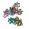
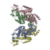
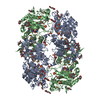
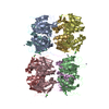
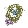
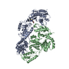
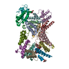
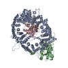
| タイトル | Structural insights into the chaperone activity of Hsp40: DnaJ binds and remodels RepE | |||||||||
 マップデータ マップデータ | 3D Resonstruction of DnaJ:RepE(1-144) complex | |||||||||
 試料 試料 |
| |||||||||
 キーワード キーワード |  Hsp40 (Hsp40) / DnaJ (Hsp40) / RepE / chaperones / protein folding (フォールディング) / electron microscopy (電子顕微鏡) Hsp40 (Hsp40) / DnaJ (Hsp40) / RepE / chaperones / protein folding (フォールディング) / electron microscopy (電子顕微鏡) | |||||||||
| 機能・相同性 |  機能・相同性情報 機能・相同性情報sigma factor antagonist activity / protein disulfide isomerase activity / chaperone cofactor-dependent protein refolding / protein unfolding / protein-disulfide reductase activity / heat shock protein binding / viral process / unfolded protein binding / フォールディング / response to heat ...sigma factor antagonist activity / protein disulfide isomerase activity / chaperone cofactor-dependent protein refolding / protein unfolding / protein-disulfide reductase activity / heat shock protein binding / viral process / unfolded protein binding / フォールディング / response to heat / protein-folding chaperone binding / protein refolding / protein-containing complex assembly / DNA複製 / protein homodimerization activity / protein-containing complex / zinc ion binding / ATP binding / 生体膜 / 細胞質基質 / 細胞質類似検索 - 分子機能 | |||||||||
| 生物種 |  Escherichia coli (大腸菌) Escherichia coli (大腸菌) | |||||||||
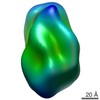
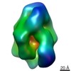
| 手法 | 単粒子再構成法 / ネガティブ染色法 / 解像度: 20.0 Å | |||||||||
 データ登録者 データ登録者 | Cuellar J / Perales-Calvo J / Muga A / Valpuesta JM / Moro F | |||||||||
 引用 引用 | ジャーナル: J Biol Chem / 年: 2013 タイトル: Structural insights into the chaperone activity of the 40-kDa heat shock protein DnaJ: binding and remodeling of a native substrate. 著者: Jorge Cuéllar / Judit Perales-Calvo / Arturo Muga / José María Valpuesta / Fernando Moro /  要旨: Hsp40 chaperones bind and transfer substrate proteins to Hsp70s and regulate their ATPase activity. The interaction of Hsp40s with native proteins modifies their structure and function. A good model ...Hsp40 chaperones bind and transfer substrate proteins to Hsp70s and regulate their ATPase activity. The interaction of Hsp40s with native proteins modifies their structure and function. A good model for this function is DnaJ, the bacterial Hsp40 that interacts with RepE, the repressor/activator of plasmid F replication, and together with DnaK regulates its function. We characterize here the structure of the DnaJ-RepE complex by electron microscopy, the first described structure of a complex between an Hsp40 and a client protein. The comparison of the complexes of DnaJ with two RepE mutants reveals an intrinsic plasticity of the DnaJ dimer that allows the chaperone to adapt to different substrates. We also show that DnaJ induces conformational changes in dimeric RepE, which increase the intermonomeric distance and remodel both RepE domains enhancing its affinity for DNA. | |||||||||
| 履歴 |
|
- 構造の表示
構造の表示
| ムービー |
ムービービューア |
|---|---|
| 構造ビューア | EMマップ: SurfViewMolmilJmol/JSmol |
| 添付画像 |

- ダウンロードとリンク
-EMDBアーカイブ
| マップデータ | emd_2333.map.gz | 728.6 KB | EMDBマップデータ形式 | |
|---|---|---|---|---|
| ヘッダ (付随情報) | emd-2333-v30.xmlemd-2333.xml | 10.4 KB 10.4 KB | 表示 表示 | EMDBヘッダ |
| 画像 | 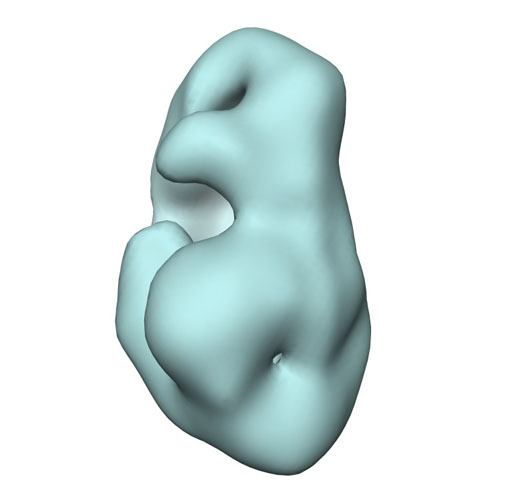 emd2333.jpg emd2333.jpg | 32.5 KB | ||
| アーカイブディレクトリ |  http://ftp.pdbj.org/pub/emdb/structures/EMD-2333ftp://ftp.pdbj.org/pub/emdb/structures/EMD-2333 http://ftp.pdbj.org/pub/emdb/structures/EMD-2333ftp://ftp.pdbj.org/pub/emdb/structures/EMD-2333 | HTTPS FTP |
-関連構造データ
-リンク
| EMDBのページ | EMDB (EBI/PDBe) / EMDataResource |
|---|---|
| 「今月の分子」の関連する項目 |


-マップ
| ファイル | ダウンロード / ファイル: emd_2333.map.gz / 形式: CCP4 / 大きさ: 1.9 MB / タイプ: IMAGE STORED AS FLOATING POINT NUMBER (4 BYTES) | ||||||||||||||||||||||||||||||||||||||||||||||||||||||||||||||||||||
|---|---|---|---|---|---|---|---|---|---|---|---|---|---|---|---|---|---|---|---|---|---|---|---|---|---|---|---|---|---|---|---|---|---|---|---|---|---|---|---|---|---|---|---|---|---|---|---|---|---|---|---|---|---|---|---|---|---|---|---|---|---|---|---|---|---|---|---|---|---|
| 注釈 | 3D Resonstruction of DnaJ:RepE(1-144) complex | ||||||||||||||||||||||||||||||||||||||||||||||||||||||||||||||||||||
| ボクセルのサイズ | X=Y=Z: 2.33 Å | ||||||||||||||||||||||||||||||||||||||||||||||||||||||||||||||||||||
| 密度 |
| ||||||||||||||||||||||||||||||||||||||||||||||||||||||||||||||||||||
| 対称性 | 空間群: 1 | ||||||||||||||||||||||||||||||||||||||||||||||||||||||||||||||||||||
| 詳細 | EMDB XML:
CCP4マップ ヘッダ情報:
| ||||||||||||||||||||||||||||||||||||||||||||||||||||||||||||||||||||
-添付データ
- 試料の構成要素
-全体 : DnaJ:RepE(1-144) complex
| 全体 | 名称: DnaJ:RepE(1-144) complex |
|---|---|
| 要素 |
|
-超分子 #1000: DnaJ:RepE(1-144) complex
| 超分子 | 名称: DnaJ:RepE(1-144) complex / タイプ: sample / ID: 1000 / 集合状態: One dimer of DnaJ binds to one dimer of RepE / Number unique components: 2 |
|---|---|
| 分子量 | 実験値: 115 KDa / 理論値: 115 KDa / 手法: Analytical ultracentrifugation (AU) |
-分子 #1: Hsp40
| 分子 | 名称: Hsp40 / タイプ: protein_or_peptide / ID: 1 / Name.synonym: DnaJ / コピー数: 2 / 集合状態: dimer / 組換発現: Yes |
|---|---|
| 由来(天然) | 生物種: Escherichia coli (大腸菌) / 細胞中の位置: cytoplasm |
| 分子量 | 実験値: 80 KDa / 理論値: 80 KDa |
| 組換発現 | 生物種: Escherichia coli (大腸菌) |
| 配列 | UniProtKB: Chaperone protein DnaJ |
-分子 #2: Shorter version of RepE (RepE 1-144)
| 分子 | 名称: Shorter version of RepE (RepE 1-144) / タイプ: protein_or_peptide / ID: 2 / Name.synonym: RepE(1-154) / コピー数: 2 / 集合状態: dimer / 組換発現: Yes |
|---|---|
| 由来(天然) | 生物種: Escherichia coli (大腸菌) / 細胞中の位置: cytoplasm |
| 分子量 | 実験値: 32 KDa / 理論値: 32 KDa |
| 組換発現 | 生物種: Escherichia coli (大腸菌) |
-実験情報
-構造解析
| 手法 | ネガティブ染色法 |
|---|---|
 解析 解析 | 単粒子再構成法 |
| 試料の集合状態 | particle |
-試料調製
| 濃度 | 0.4 mg/mL |
|---|---|
| 緩衝液 | pH: 7.4 / 詳細: 20mM Hepes pH 7.4, 50mM KCl, 5mM DTT and 0.1mM EDTA |
| 染色 | タイプ: NEGATIVE 詳細: Samples (either DnaJ:RepE, DnaJ:RepE1-144 or DnaJ:RepE54 complexes) were diluted 1:100 in 20mM Hepes pH 7.4, 50mM KCl, 5mM DTT and 0.1mM EDTA buffer, applied onto carbon-coated copper grids ...詳細: Samples (either DnaJ:RepE, DnaJ:RepE1-144 or DnaJ:RepE54 complexes) were diluted 1:100 in 20mM Hepes pH 7.4, 50mM KCl, 5mM DTT and 0.1mM EDTA buffer, applied onto carbon-coated copper grids and stained with 2% uranyl acetate. |
| グリッド | 詳細: 300 mesh Cu/Rh grid with thin carbon support and glow discharged |
| 凍結 | 凍結剤: NONE / 装置: OTHER |
- 電子顕微鏡法
| 顕微鏡 | JEOL 1200EXII |
|---|---|
| 電子線 | 加速電圧: 100 kV / 電子線源: TUNGSTEN HAIRPIN |
| 電子光学系 | 照射モード: FLOOD BEAM / 撮影モード: BRIGHT FIELDBright-field microscopy / Cs: 5.6 mm / 倍率(公称値): 60000 |
| 試料ステージ | 試料ホルダー: Standard Jeol 1200 holder / 試料ホルダーモデル: JEOL |
| アライメント法 | Legacy - 非点収差: Objective lens astigmatism was corrected at 100,000 times magnification |
| 詳細 | Additional details about microscope model:JEOL 1200EXII |
| 日付 | 2011年6月8日 |
| 撮影 | カテゴリ: FILM / フィルム・検出器のモデル: KODAK SO-163 FILM / デジタル化 - スキャナー: ZEISS SCAI / デジタル化 - サンプリング間隔: 14 µm / 実像数: 270 / 平均電子線量: 10 e/Å2 / ビット/ピクセル: 8 |
-画像解析
| CTF補正 | 詳細: CTFFIND |
|---|---|
| 最終 2次元分類 | クラス数: 75 |
| 最終 再構成 | 想定した対称性 - 点群: C1 (非対称) / アルゴリズム: OTHER / 解像度のタイプ: BY AUTHOR / 解像度: 20.0 Å / 解像度の算出法: FSC 0.5 CUT-OFF / ソフトウェア - 名称: EMAN, XMIPP / 使用した粒子像数: 9623 |
| 詳細 | The particles were selected by manual picking |
