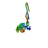
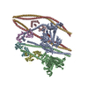
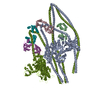
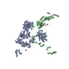
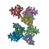
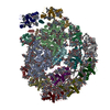
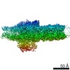
ジャーナル: Sci Adv / 年: 2021 タイトル: Cryo-EM structure of the autoinhibited state of myosin-2. 著者: Sarah M Heissler / Amandeep S Arora / Neil Billington / James R Sellers / Krishna Chinthalapudi / 要旨: We solved the near-atomic resolution structure of smooth muscle myosin-2 in the autoinhibited state (10) using single-particle cryo–electron microscopy. The 3.4-Å structure reveals the precise ...We solved the near-atomic resolution structure of smooth muscle myosin-2 in the autoinhibited state (10) using single-particle cryo–electron microscopy. The 3.4-Å structure reveals the precise molecular architecture of 10 and the structural basis for myosin-2 regulation. We reveal the position of the phosphorylation sites that control myosin autoinhibition and activation by phosphorylation of the regulatory light chain. Further, we present a previously unidentified conformational state in myosin-2 that traps ADP and P produced by the hydrolysis of ATP in the active site. This noncanonical state represents a branch of the myosin enzyme cycle and explains the autoinhibition of the enzyme function of 10 along with its reduced affinity for actin. Together, our structure defines the molecular mechanisms that drive 10 formation, stabilization, and relief by phosphorylation of the regulatory light chain.
 ムービー
ムービー コントローラー


 データを開く
 基本情報
基本情報 要素
要素 キーワード
キーワード CONTRACTILE PROTEIN /
  機能・相同性情報
 データ登録者
 引用
引用
 構造の表示
構造の表示 ダウンロードとリンク
 その他のダウンロード
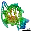

 PDBj
PDBj


 集合体
集合体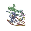


 分子量: 427.201 Da / 分子数: 2 / 由来タイプ: 合成 / 式: C10H15N5O10P2 / コメント: ADP, エネルギー貯蔵分子*YM
 分子量: 94.971 Da / 分子数: 2 / 由来タイプ: 合成 / 式: PO4
 分子量: 24.305 Da / 分子数: 4 / 由来タイプ: 合成 / 式: Mg
 試料調製
試料調製 電子顕微鏡撮影
 解析
解析