 ムービー
ムービー コントローラー


+ データを開く

- 基本情報
基本情報
| 登録情報 | データベース: PDB / ID: 6v4k | |||||||||
|---|---|---|---|---|---|---|---|---|---|---|
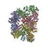
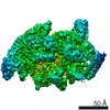
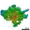
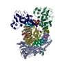
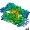
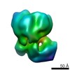
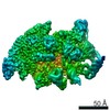
| タイトル | Structure of TrkH-TrkA in complex with ADP | |||||||||
 要素 要素 |
| |||||||||
 キーワード キーワード |  TRANSPORT PROTEIN (運搬体タンパク質) / ion channel (イオンチャネル) / TrkH / TrkA / nucleotide binding (ヌクレオチド) TRANSPORT PROTEIN (運搬体タンパク質) / ion channel (イオンチャネル) / TrkH / TrkA / nucleotide binding (ヌクレオチド) | |||||||||
| 機能・相同性 |  機能・相同性情報 機能・相同性情報potassium ion transmembrane transporter activity / potassium:chloride symporter activity / potassium ion binding / potassium channel activity / potassium ion transmembrane transport / membrane => GO:0016020 / nucleotide binding / protein homodimerization activity / identical protein binding / metal ion binding / 細胞膜類似検索 - 分子機能 | |||||||||
| 生物種 |   Vibrio parahaemolyticus (腸炎ビブリオ) Vibrio parahaemolyticus (腸炎ビブリオ) | |||||||||
| 手法 | X線回折 / シンクロトロン / 分子置換 / 解像度: 3.53004144504 Å | |||||||||
 データ登録者 データ登録者 | Zhou, M. / Zhang, H. | |||||||||
| 資金援助 |  米国, 2件 米国, 2件
| |||||||||
 引用 引用 | ジャーナル: Nat Commun / 年: 2020 タイトル: TrkA undergoes a tetramer-to-dimer conversion to open TrkH which enables changes in membrane potential. 著者: Hanzhi Zhang / Yaping Pan / Liya Hu / M Ashley Hudson / Katrina S Hofstetter / Zhichun Xu / Mingqiang Rong / Zhao Wang / B V Venkataram Prasad / Steve W Lockless / Wah Chiu / Ming Zhou / 要旨: TrkH is a bacterial ion channel implicated in K uptake and pH regulation. TrkH assembles with its regulatory protein, TrkA, which closes the channel when bound to ADP and opens it when bound to ATP. ...TrkH is a bacterial ion channel implicated in K uptake and pH regulation. TrkH assembles with its regulatory protein, TrkA, which closes the channel when bound to ADP and opens it when bound to ATP. However, it is unknown how nucleotides control the gating of TrkH through TrkA. Here we report the structures of the TrkH-TrkA complex in the presence of ADP or ATP. TrkA forms a tetrameric ring when bound to ADP and constrains TrkH to a closed conformation. The TrkA ring splits into two TrkA dimers in the presence of ATP and releases the constraints on TrkH, resulting in an open channel conformation. Functional studies show that both the tetramer-to-dimer conversion of TrkA and the loss of constraints on TrkH are required for channel gating. In addition, deletion of TrkA in Escherichia coli depolarizes the cell, suggesting that the TrkH-TrkA complex couples changes in intracellular nucleotides to membrane potential. | |||||||||
| 履歴 |
|
- 構造の表示
構造の表示
| 構造ビューア | 分子: MolmilJmol/JSmol |
|---|
- ダウンロードとリンク
-ダウンロード
| PDBx/mmCIF形式 | 6v4k.cif.gz | 1.4 MB | 表示 | PDBx/mmCIF形式 |
|---|---|---|---|---|
| PDB形式 | pdb6v4k.ent.gz | 1.2 MB | 表示 | PDB形式 |
| PDBx/mmJSON形式 | 6v4k.json.gz | ツリー表示 | PDBx/mmJSON形式 | |
| その他 |  その他のダウンロード その他のダウンロード |
-検証レポート
| アーカイブディレクトリ | https://data.pdbj.org/pub/pdb/validation_reports/v4/6v4kftp://data.pdbj.org/pub/pdb/validation_reports/v4/6v4k | HTTPS FTP |
|---|
-関連構造データ


-リンク
 PDBj
PDBj
- 集合体
集合体
| 登録構造単位 | 
| ||||||||||||
|---|---|---|---|---|---|---|---|---|---|---|---|---|---|
| 1 |
| ||||||||||||
| 単位格子 |
|
-要素
| #1: タンパク質 | 分子量: 53104.375 Da / 分子数: 4 / 由来タイプ: 組換発現 由来: (組換発現) Vibrio parahaemolyticus (腸炎ビブリオ)遺伝子: ACS91_07345, BA740_20060, BS585_02545, C1S91_23870, C9I78_16125, CA163_10465, CGH73_23020, CGJ02_21120, FHP20_22005, WR32_21470 発現宿主: Escherichia coli BL21(DE3) (大腸菌) / 参照: UniProt: A0A0D1QU68, UniProt: Q87TN7*PLUS#2: タンパク質 | 分子量: 50193.086 Da / 分子数: 4 / 由来タイプ: 組換発現 由来: (組換発現) Vibrio parahaemolyticus (腸炎ビブリオ)遺伝子: trkA, sapG, ACS91_07035, BA740_20405, C1S91_23530, CGH73_23350, CGJ02_20785, WR32_21780 発現宿主: Escherichia coli BL21(DE3) (大腸菌) / 参照: UniProt: A0A072LGS4, UniProt: Q87KD2*PLUS#3: 化合物 | ChemComp-ADP / アデノシン二リン酸  分子量: 427.201 Da / 分子数: 4 / 由来タイプ: 合成 / 式: C10H15N5O10P2 / コメント: ADP, エネルギー貯蔵分子*YM 分子量: 427.201 Da / 分子数: 4 / 由来タイプ: 合成 / 式: C10H15N5O10P2 / コメント: ADP, エネルギー貯蔵分子*YM研究の焦点であるリガンドがあるか | N | |
|---|
-実験情報
-実験
| 実験 | 手法: X線回折 / 使用した結晶の数: 1 |
|---|
- 試料調製
試料調製
| 結晶 | マシュー密度: 4.1 Å3/Da / 溶媒含有率: 69.98 % |
|---|---|
| 結晶化 | 温度: 293 K / 手法: 蒸気拡散法, シッティングドロップ法 / pH: 7.2 / 詳細: 100 mM HEPES, pH 7.2, 26% PEG400, 10% 2-propanol |
-データ収集
| 回折 | 平均測定温度: 80 K / Serial crystal experiment: N |
|---|---|
| 放射光源 | 由来: シンクロトロン / サイト: APS / ビームライン: 24-ID-C / 波長: 1 Å |
| 検出器 | タイプ: DECTRIS PILATUS 6M-F / 検出器: PIXEL / 日付: 2017年7月30日 |
| 放射 | モノクロメーター: cryo-cooled double crystal Si(111) プロトコル: SINGLE WAVELENGTH / 単色(M)・ラウエ(L): M / 散乱光タイプ: x-ray |
| 放射波長 | 波長: 1 Å / 相対比: 1 |
| 反射 | 解像度: 3.5→50 Å / Num. obs: 81759 / % possible obs: 99.3 % / 冗長度: 6.8 % / Biso Wilson estimate: 21.3655473934 Å2 / Rmerge(I) obs: 0.065 / Net I/σ(I): 30.63 |
| 反射 シェル | 解像度: 3.52→3.58 Å / Rmerge(I) obs: 1.7 / Num. unique obs: 4081 |
- 解析
解析
| ソフトウェア |
| |||||||||||||||||||||||||||||||||||||||||||||||||||||||||||||||||||||||||||||||||||||||||||||||||||||||||||||||||||||||||||||||||||||||||||||||||||||||||||||||||
|---|---|---|---|---|---|---|---|---|---|---|---|---|---|---|---|---|---|---|---|---|---|---|---|---|---|---|---|---|---|---|---|---|---|---|---|---|---|---|---|---|---|---|---|---|---|---|---|---|---|---|---|---|---|---|---|---|---|---|---|---|---|---|---|---|---|---|---|---|---|---|---|---|---|---|---|---|---|---|---|---|---|---|---|---|---|---|---|---|---|---|---|---|---|---|---|---|---|---|---|---|---|---|---|---|---|---|---|---|---|---|---|---|---|---|---|---|---|---|---|---|---|---|---|---|---|---|---|---|---|---|---|---|---|---|---|---|---|---|---|---|---|---|---|---|---|---|---|---|---|---|---|---|---|---|---|---|---|---|---|---|---|---|
| 精密化 | 構造決定の手法: 分子置換 開始モデル: PDB entry 4J9U 解像度: 3.53004144504→49.8219581438 Å / SU ML: 0.370948467346 / 交差検証法: FREE R-VALUE / σ(F): 1.33788131981 / 位相誤差: 27.9833370918
| |||||||||||||||||||||||||||||||||||||||||||||||||||||||||||||||||||||||||||||||||||||||||||||||||||||||||||||||||||||||||||||||||||||||||||||||||||||||||||||||||
| 溶媒の処理 | 減衰半径: 0.9 Å / VDWプローブ半径: 1.11 Å | |||||||||||||||||||||||||||||||||||||||||||||||||||||||||||||||||||||||||||||||||||||||||||||||||||||||||||||||||||||||||||||||||||||||||||||||||||||||||||||||||
| 原子変位パラメータ | Biso mean: 80.3680566079 Å2 | |||||||||||||||||||||||||||||||||||||||||||||||||||||||||||||||||||||||||||||||||||||||||||||||||||||||||||||||||||||||||||||||||||||||||||||||||||||||||||||||||
| 精密化ステップ | サイクル: LAST / 解像度: 3.53004144504→49.8219581438 Å
| |||||||||||||||||||||||||||||||||||||||||||||||||||||||||||||||||||||||||||||||||||||||||||||||||||||||||||||||||||||||||||||||||||||||||||||||||||||||||||||||||
| 拘束条件 |
| |||||||||||||||||||||||||||||||||||||||||||||||||||||||||||||||||||||||||||||||||||||||||||||||||||||||||||||||||||||||||||||||||||||||||||||||||||||||||||||||||
| LS精密化 シェル |
|
