Ministry of Education, Culture, Sports, Science and Technology (Japan)
jp15076210
日本
Ministry of Education, Culture, Sports, Science and Technology (Japan)
jp20050030
日本
Ministry of Education, Culture, Sports, Science and Technology (Japan)
jp22018027
日本
Ministry of Education, Culture, Sports, Science and Technology (Japan)
jp23120525
日本
Ministry of Education, Culture, Sports, Science and Technology (Japan)
jp25120725
日本
Ministry of Education, Culture, Sports, Science and Technology (Japan)
jp15H01647
日本
Ministry of Education, Culture, Sports, Science and Technology (Japan)
jp17H05891
日本
Ministry of Education, Culture, Sports, Science and Technology (Japan)
jp26104535
日本
引用
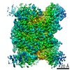
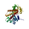
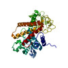
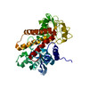
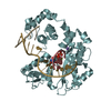
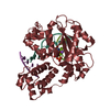
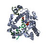
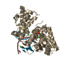
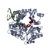
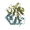
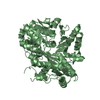
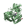
ジャーナル: FEBS J / 年: 2020 タイトル: Energy landscape of domain motion in glutamate dehydrogenase deduced from cryo-electron microscopy. 著者: Mao Oide / Takayuki Kato / Tomotaka Oroguchi / Masayoshi Nakasako / 要旨: Analysis of the conformational changes of protein is important to elucidate the mechanisms of protein motions correlating with their function. Here, we studied the spontaneous domain motion of ...Analysis of the conformational changes of protein is important to elucidate the mechanisms of protein motions correlating with their function. Here, we studied the spontaneous domain motion of unliganded glutamate dehydrogenase from Thermococcus profundus using cryo-electron microscopy and proposed a novel method to construct free-energy landscape of protein conformations. Each subunit of the homo-hexameric enzyme comprises nucleotide-binding domain (NAD domain) and hexamer-forming core domain. A large active-site cleft is situated between the two domains and varies from open to close according to the motion of a NAD domain. A three-dimensional map reconstructed from all cryo-electron microscopy images displayed disordered volumes of NAD domains, suggesting that NAD domains in the collected images adopted various conformations in domain motion. Focused classifications on NAD domain of subunits provided several maps of possible conformations in domain motion. To deduce what kinds of conformations appeared in EM images, we developed a novel analysis method that describe the EM maps as a linear combination of representative conformations appearing in a 200-ns molecular dynamics simulation as reference. The analysis enabled us to estimate the appearance frequencies of the representative conformations, which illustrated a free-energy landscape in domain motion. In the open/close domain motion, two free-energy basins hindered the direct transformation from open to closed state. Structure models constructed for representative EM maps in classifications demonstrated the correlation between the energy landscape and conformations in domain motion. Based on the results, the domain motion in glutamate dehydrogenase and the analysis method to visualize conformational changes and free-energy landscape were discussed. DATABASE: The EM maps of the four conformations were deposited to Electron Microscopy Data Bank (EMDB) as accession codes EMD-9845 (open), EMD-9846 (half-open1), EMD-9847 (half-open2), and EMD-9848 (closed), respectively. In addition, the structural models built for the four conformations were deposited to the Protein Data Bank (PDB) as accession codes 6JN9 (open), 6JNA (half-open1), 6JNC (half-open2), and 6JND (closed), respectively.
 ムービー
ムービー コントローラー


 データを開く
 基本情報
基本情報 要素
要素 グルタミン酸デヒドロゲナーゼ
  キーワード
キーワード 機能・相同性情報

 データ登録者
 日本, 14件
  引用
引用 構造の表示
構造の表示 ダウンロードとリンク
 その他のダウンロード


 PDBj
PDBj 集合体
集合体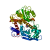


 分子量: 122.143 Da / 分子数: 1 / 由来タイプ: 合成 / 式: C4H12NO3 / コメント: pH緩衝剤*YM
 試料調製
試料調製 電子顕微鏡撮影
 解析
解析