 ムービー
ムービー コントローラー


+ データを開く

- 基本情報
基本情報
| 登録情報 | データベース: PDB / ID: 6hwi | ||||||||||||||||||||||||||||||
|---|---|---|---|---|---|---|---|---|---|---|---|---|---|---|---|---|---|---|---|---|---|---|---|---|---|---|---|---|---|---|---|
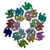
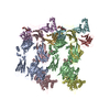
| タイトル | Immature M-PMV capsid hexamer structure in intact virus particles | ||||||||||||||||||||||||||||||
 要素 要素 | Gag-Pro-Pol polyprotein | ||||||||||||||||||||||||||||||
 キーワード キーワード |  VIRAL PROTEIN (ウイルスタンパク質) / M-PMV / capsid (カプシド) / hexamer (オリゴマー) VIRAL PROTEIN (ウイルスタンパク質) / M-PMV / capsid (カプシド) / hexamer (オリゴマー) | ||||||||||||||||||||||||||||||
| 機能・相同性 |  機能・相同性情報dUTP diphosphatase / dUTP diphosphatase activity / nucleotide metabolic process / 加水分解酵素; プロテアーゼ; ペプチド結合加水分解酵素; アスパラギン酸プロテアーゼ / リボヌクレアーゼH / viral process / DNA integration / 逆転写酵素 / viral genome integration into host DNA / establishment of integrated proviral latency ...dUTP diphosphatase / dUTP diphosphatase activity / nucleotide metabolic process / 加水分解酵素; プロテアーゼ; ペプチド結合加水分解酵素; アスパラギン酸プロテアーゼ / リボヌクレアーゼH / viral process / DNA integration / 逆転写酵素 / viral genome integration into host DNA / establishment of integrated proviral latency / RNA-directed DNA polymerase activity / 転移酵素; リンを含む基を移すもの; 核酸を移すもの / RNA-DNA hybrid ribonuclease activity / viral nucleocapsid / DNA recombination / structural constituent of virion / 加水分解酵素; エステル加水分解酵素 / DNAポリメラーゼ / aspartic-type endopeptidase activity / DNA-directed DNA polymerase activity / symbiont entry into host cell / タンパク質分解 / DNA binding / RNA binding / zinc ion binding 機能・相同性情報dUTP diphosphatase / dUTP diphosphatase activity / nucleotide metabolic process / 加水分解酵素; プロテアーゼ; ペプチド結合加水分解酵素; アスパラギン酸プロテアーゼ / リボヌクレアーゼH / viral process / DNA integration / 逆転写酵素 / viral genome integration into host DNA / establishment of integrated proviral latency ...dUTP diphosphatase / dUTP diphosphatase activity / nucleotide metabolic process / 加水分解酵素; プロテアーゼ; ペプチド結合加水分解酵素; アスパラギン酸プロテアーゼ / リボヌクレアーゼH / viral process / DNA integration / 逆転写酵素 / viral genome integration into host DNA / establishment of integrated proviral latency / RNA-directed DNA polymerase activity / 転移酵素; リンを含む基を移すもの; 核酸を移すもの / RNA-DNA hybrid ribonuclease activity / viral nucleocapsid / DNA recombination / structural constituent of virion / 加水分解酵素; エステル加水分解酵素 / DNAポリメラーゼ / aspartic-type endopeptidase activity / DNA-directed DNA polymerase activity / symbiont entry into host cell / タンパク質分解 / DNA binding / RNA binding / zinc ion binding類似検索 - 分子機能 | ||||||||||||||||||||||||||||||
| 生物種 |  Mason-Pfizer monkey virus (ウイルス) Mason-Pfizer monkey virus (ウイルス) | ||||||||||||||||||||||||||||||
| 手法 | 電子顕微鏡法 / サブトモグラム平均法 / クライオ電子顕微鏡法 / 解像度: 7.2 Å | ||||||||||||||||||||||||||||||
 データ登録者 データ登録者 | Qu, K. / Glass, B. / Dolezal, M. / Schur, F.K.M. / Rein, A. / Rumlova, M. / Ruml, T. / Kraeusslich, H.G. / Briggs, J.A.G. | ||||||||||||||||||||||||||||||
| 資金援助 |  ドイツ, ドイツ,  英国, 英国,  チェコ, 9件 チェコ, 9件
| ||||||||||||||||||||||||||||||
 引用 引用 | ジャーナル: Proc Natl Acad Sci U S A / 年: 2018 タイトル: Structure and architecture of immature and mature murine leukemia virus capsids. 著者: Kun Qu / Bärbel Glass / Michal Doležal / Florian K M Schur / Brice Murciano / Alan Rein / Michaela Rumlová / Tomáš Ruml / Hans-Georg Kräusslich / John A G Briggs /   要旨: Retroviruses assemble and bud from infected cells in an immature form and require proteolytic maturation for infectivity. The CA (capsid) domains of the Gag polyproteins assemble a protein lattice as ...Retroviruses assemble and bud from infected cells in an immature form and require proteolytic maturation for infectivity. The CA (capsid) domains of the Gag polyproteins assemble a protein lattice as a truncated sphere in the immature virion. Proteolytic cleavage of Gag induces dramatic structural rearrangements; a subset of cleaved CA subsequently assembles into the mature core, whose architecture varies among retroviruses. Murine leukemia virus (MLV) is the prototypical γ-retrovirus and serves as the basis of retroviral vectors, but the structure of the MLV CA layer is unknown. Here we have combined X-ray crystallography with cryoelectron tomography to determine the structures of immature and mature MLV CA layers within authentic viral particles. This reveals the structural changes associated with maturation, and, by comparison with HIV-1, uncovers conserved and variable features. In contrast to HIV-1, most MLV CA is used for assembly of the mature core, which adopts variable, multilayered morphologies and does not form a closed structure. Unlike in HIV-1, there is similarity between protein-protein interfaces in the immature MLV CA layer and those in the mature CA layer, and structural maturation of MLV could be achieved through domain rotations that largely maintain hexameric interactions. Nevertheless, the dramatic architectural change on maturation indicates that extensive disassembly and reassembly are required for mature core growth. The core morphology suggests that wrapping of the genome in CA sheets may be sufficient to protect the MLV ribonucleoprotein during cell entry. | ||||||||||||||||||||||||||||||
| 履歴 |
|
- 構造の表示
構造の表示
| ムービー |
ムービービューア |
|---|---|
| 構造ビューア | 分子: MolmilJmol/JSmol |
- ダウンロードとリンク
-ダウンロード
| PDBx/mmCIF形式 | 6hwi.cif.gz | 85.9 KB | 表示 | PDBx/mmCIF形式 |
|---|---|---|---|---|
| PDB形式 | pdb6hwi.ent.gz | 58.6 KB | 表示 | PDB形式 |
| PDBx/mmJSON形式 | 6hwi.json.gz | ツリー表示 | PDBx/mmJSON形式 | |
| その他 |  その他のダウンロード その他のダウンロード |
-検証レポート
| アーカイブディレクトリ | https://data.pdbj.org/pub/pdb/validation_reports/hw/6hwiftp://data.pdbj.org/pub/pdb/validation_reports/hw/6hwi | HTTPS FTP |
|---|
-関連構造データ
| 関連構造データ | 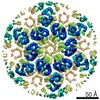 0290MC 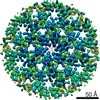 0291C 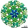 0292C 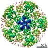 0293C  4419C  4421C  4422C 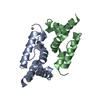 6gzaC 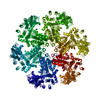 6hwwC 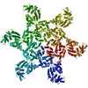 6hwxC 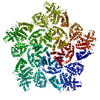 6hwyC M: このデータのモデリングに利用したマップデータ C: 同じ文献を引用 ( |
|---|---|
| 類似構造データ |


-リンク
 PDBj
PDBj


- 集合体
集合体
| 登録構造単位 | 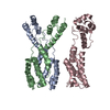
|
|---|---|
| 1 | x 6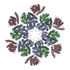
|
-要素
| #1: タンパク質 | 分子量: 21817.359 Da / 分子数: 3 / 由来タイプ: 組換発現 由来: (組換発現) Mason-Pfizer monkey virus (ウイルス)遺伝子: gag-pro-pol / 発現宿主:  Homo sapiens (ヒト) / 参照: UniProt: P07572, UniProt: P07570*PLUS Homo sapiens (ヒト) / 参照: UniProt: P07572, UniProt: P07570*PLUS |
|---|
-実験情報
-実験
| 実験 | 手法: 電子顕微鏡法 |
|---|---|
| EM実験 | 試料の集合状態: PARTICLE / 3次元再構成法: サブトモグラム平均法 |
- 試料調製
試料調製
| 構成要素 | 名称: Mason-Pfizer monkey virus / タイプ: VIRUS / Entity ID: all / 由来: RECOMBINANT |
|---|---|
| 由来(天然) | 生物種: Mason-Pfizer monkey virus (ウイルス) |
| 由来(組換発現) | 生物種: Homo sapiens (ヒト) / 細胞: HEK 293T / プラスミド: pSHRM15 D26N |
| ウイルスについての詳細 | 中空か: NO / エンベロープを持つか: YES / 単離: STRAIN / タイプ: VIRION |
| 緩衝液 | pH: 6 |
| 試料 | 包埋: NO / シャドウイング: NO / 染色: NO / 凍結: YES 詳細: Purified virus solution was inactivated and diluted 1:1 with PBS containing 10 nm colloidal gold. |
| 試料支持 | グリッドの材料: COPPER / グリッドのサイズ: 300 divisions/in. / グリッドのタイプ: C-flat-2/1 |
| 急速凍結 | 装置: FEI VITROBOT MARK II / 凍結剤: ETHANE / 湿度: 95 % / 凍結前の試料温度: 288 K |
- 電子顕微鏡撮影
| 実験機器 |  モデル: Titan Krios / 画像提供: FEI Company |
|---|---|
| 顕微鏡 | モデル: FEI TITAN KRIOS |
| 電子銃 | 電子線源: FIELD EMISSION GUN / 加速電圧: 300 kV / 照射モード: FLOOD BEAM |
| 電子レンズ | モード: BRIGHT FIELDBright-field microscopy / 倍率(公称値): 105000 X / 最大 デフォーカス(公称値): 4500 nm / 最小 デフォーカス(公称値): 2000 nm / Cs: 2.7 mm / C2レンズ絞り径: 50 µm / アライメント法: ZEMLIN TABLEAU |
| 試料ホルダ | 凍結剤: NITROGEN 試料ホルダーモデル: FEI TITAN KRIOS AUTOGRID HOLDER |
| 撮影 | 平均露光時間: 0.6 sec. / 電子線照射量: 2 e/Å2 / 検出モード: SUPER-RESOLUTION フィルム・検出器のモデル: GATAN K2 QUANTUM (4k x 4k) 撮影したグリッド数: 1 詳細: Dose fluctuation was caused by the ring collapse of FEG during data collection. |
| 電子光学装置 | エネルギーフィルター名称: GIF Quantum LS / エネルギーフィルタースリット幅: 20 eV |
| 画像スキャン | サンプリングサイズ: 5 µm / 横: 3710 / 縦: 3838 / 動画フレーム数/画像: 6 / 利用したフレーム数/画像: 1-6 |
- 解析
解析
| EMソフトウェア |
| ||||||||||||||||||||||||||||||||||||||||||||||||||||
|---|---|---|---|---|---|---|---|---|---|---|---|---|---|---|---|---|---|---|---|---|---|---|---|---|---|---|---|---|---|---|---|---|---|---|---|---|---|---|---|---|---|---|---|---|---|---|---|---|---|---|---|---|---|
| CTF補正 | タイプ: PHASE FLIPPING ONLY | ||||||||||||||||||||||||||||||||||||||||||||||||||||
| 対称性 | 点対称性: C6 (6回回転対称) | ||||||||||||||||||||||||||||||||||||||||||||||||||||
| 3次元再構成 | 解像度: 7.2 Å / 解像度の算出法: FSC 0.143 CUT-OFF / 粒子像の数: 17109 / 対称性のタイプ: POINT | ||||||||||||||||||||||||||||||||||||||||||||||||||||
| EM volume selection | Num. of tomograms: 34 / Num. of volumes extracted: 17109 | ||||||||||||||||||||||||||||||||||||||||||||||||||||
| 原子モデル構築 | プロトコル: FLEXIBLE FIT / 空間: REAL |
