National Institutes of Health/National Institute of General Medical Sciences (NIH/NIGMS)
GM24485
米国
引用
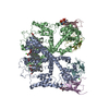
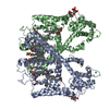
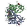
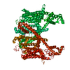
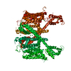
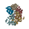
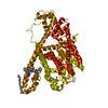
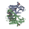
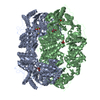
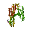
ジャーナル: Proc Natl Acad Sci U S A / 年: 2018 タイトル: Structural basis for activation of voltage sensor domains in an ion channel TPC1. 著者: Alexander F Kintzer / Evan M Green / Pawel K Dominik / Michael Bridges / Jean-Paul Armache / Dawid Deneka / Sangwoo S Kim / Wayne Hubbell / Anthony A Kossiakoff / Yifan Cheng / Robert M Stroud / 要旨: Voltage-sensing domains (VSDs) couple changes in transmembrane electrical potential to conformational changes that regulate ion conductance through a central channel. Positively charged amino acids ...Voltage-sensing domains (VSDs) couple changes in transmembrane electrical potential to conformational changes that regulate ion conductance through a central channel. Positively charged amino acids inside each sensor cooperatively respond to changes in voltage. Our previous structure of a TPC1 channel captured an example of a resting-state VSD in an intact ion channel. To generate an activated-state VSD in the same channel we removed the luminal inhibitory Ca-binding site (Ca), which shifts voltage-dependent opening to more negative voltage and activation at 0 mV. Cryo-EM reveals two coexisting structures of the VSD, an intermediate state 1 that partially closes access to the cytoplasmic side but remains occluded on the luminal side and an intermediate activated state 2 in which the cytoplasmic solvent access to the gating charges closes, while luminal access partially opens. Activation can be thought of as moving a hydrophobic insulating region of the VSD from the external side to an alternate grouping on the internal side. This effectively moves the gating charges from the inside potential to that of the outside. Activation also requires binding of Ca to a cytoplasmic site (Ca). An X-ray structure with Ca removed and a near-atomic resolution cryo-EM structure with Ca removed define how dramatic conformational changes in the cytoplasmic domains may communicate with the VSD during activation. Together four structures provide a basis for understanding the voltage-dependent transition from resting to activated state, the tuning of VSD by thermodynamic stability, and this channel's requirement of cytoplasmic Ca ions for activation.
 ムービー
ムービー コントローラー


 データを開く
 基本情報
基本情報 要素
要素 キーワード
キーワード membrane protein/inhibitor (生体膜) /
  機能・相同性情報
 データ登録者
 米国, 1件
  引用
引用 構造の表示
構造の表示 ダウンロードとリンク
 その他のダウンロード


 PDBj
PDBj
 集合体
集合体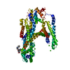


 分子量: 40.078 Da / 分子数: 5 / 由来タイプ: 組換発現 / 式: Ca
 分子量: 514.591 Da / 分子数: 1 / 由来タイプ: 組換発現 / 式: C30H31FN4O3
 試料調製
試料調製 解析
解析