National Institutes of Health/National Institute of Neurological Disorders and Stroke (NIH/NINDS)
NS083174
米国
National Institutes of Health/National Institute of Dental and Craniofacial Research (NIH/NIDCR)
DE022358
米国
Howard Hughes Medical Institute (HHMI)
米国
引用
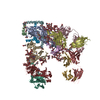
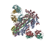
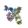
ジャーナル: Nature / 年: 2018 タイトル: Structure of the mechanically activated ion channel Piezo1. 著者: Kei Saotome / Swetha E Murthy / Jennifer M Kefauver / Tess Whitwam / Ardem Patapoutian / Andrew B Ward / 要旨: Piezo1 and Piezo2 are mechanically activated ion channels that mediate touch perception, proprioception and vascular development. Piezo proteins are distinct from other ion channels and their ...Piezo1 and Piezo2 are mechanically activated ion channels that mediate touch perception, proprioception and vascular development. Piezo proteins are distinct from other ion channels and their structure remains poorly defined, which impedes detailed study of their gating and ion permeation properties. Here we report a high-resolution cryo-electron microscopy structure of the mouse Piezo1 trimer. The detergent-solubilized complex adopts a three-bladed propeller shape with a curved transmembrane region containing at least 26 transmembrane helices per protomer. The flexible propeller blades can adopt distinct conformations, and consist of a series of four-transmembrane helical bundles that we term Piezo repeats. Carboxy-terminal domains line the central ion pore, and the channel is closed by constrictions in the cytosol. A kinked helical beam and anchor domain link the Piezo repeats to the pore, and are poised to control gating allosterically. The structure provides a foundation to dissect further how Piezo channels are regulated by mechanical force.
Piezo-typemechanosensitiveionchannelcomponent1,Piezo-typemechanosensitiveionchannelcomponent1,mousePiezo1,Piezo-typemechanosensitiveionchannelcomponent1,Piezo-typemechanosensitiveionchannelcomponent1 / Protein FAM38A
 ムービー
ムービー コントローラー


 データを開く
 基本情報
基本情報 要素
要素 キーワード
キーワード MEMBRANE PROTEIN (膜タンパク質) /
  機能・相同性情報
 データ登録者
 米国, 4件
  引用
引用 構造の表示
構造の表示 ダウンロードとリンク
 その他のダウンロード


 PDBj
PDBj
 集合体
集合体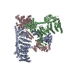

 試料調製
試料調製 電子顕微鏡撮影
 解析
解析