 ムービー
ムービー コントローラー


+ データを開く

- 基本情報
基本情報
| 登録情報 | データベース: PDB / ID: 6bno | ||||||
|---|---|---|---|---|---|---|---|
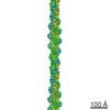
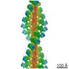
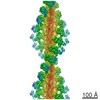
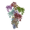
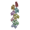
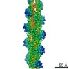
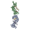
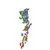
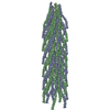
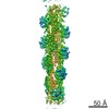
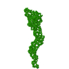
| タイトル | Structure of bare actin filament | ||||||
 要素 要素 | Actin, alpha skeletal muscle | ||||||
 キーワード キーワード | CONTRACTILE PROTEIN / Cytoskeleton (細胞骨格) / Filament | ||||||
| 機能・相同性 |  機能・相同性情報 機能・相同性情報cytoskeletal motor activator activity / tropomyosin binding / myosin heavy chain binding / mesenchyme migration / troponin I binding / actin filament bundle / filamentous actin / actin filament bundle assembly / skeletal muscle thin filament assembly / striated muscle thin filament ...cytoskeletal motor activator activity / tropomyosin binding / myosin heavy chain binding / mesenchyme migration / troponin I binding / actin filament bundle / filamentous actin / actin filament bundle assembly / skeletal muscle thin filament assembly / striated muscle thin filament / skeletal muscle myofibril / actin monomer binding / skeletal muscle fiber development / stress fiber / titin binding / actin filament polymerization / filopodium / マイクロフィラメント / 加水分解酵素; 酸無水物に作用; 酸無水物に作用・細胞または細胞小器官の運動に関与 / calcium-dependent protein binding / lamellipodium / cell body / hydrolase activity / protein domain specific binding / calcium ion binding / positive regulation of gene expression / magnesium ion binding / ATP binding / identical protein binding / 細胞質類似検索 - 分子機能 | ||||||
| 生物種 |  Oryctolagus cuniculus (ウサギ) Oryctolagus cuniculus (ウサギ) | ||||||
| 手法 | 電子顕微鏡法 / らせん対称体再構成法 / クライオ電子顕微鏡法 / 解像度: 5.5 Å | ||||||
 データ登録者 データ登録者 | Gurel, P.S. / Alushin, G.A. | ||||||
| 資金援助 |  米国, 1件 米国, 1件
| ||||||
 引用 引用 | ジャーナル: Elife / 年: 2017 タイトル: Cryo-EM structures reveal specialization at the myosin VI-actin interface and a mechanism of force sensitivity. 著者: Pinar S Gurel / Laura Y Kim / Paul V Ruijgrok / Tosan Omabegho / Zev Bryant / Gregory M Alushin / 要旨: Despite extensive scrutiny of the myosin superfamily, the lack of high-resolution structures of actin-bound states has prevented a complete description of its mechanochemical cycle and limited ...Despite extensive scrutiny of the myosin superfamily, the lack of high-resolution structures of actin-bound states has prevented a complete description of its mechanochemical cycle and limited insight into how sequence and structural diversification of the motor domain gives rise to specialized functional properties. Here we present cryo-EM structures of the unique minus-end directed myosin VI motor domain in rigor (4.6 Å) and Mg-ADP (5.5 Å) states bound to F-actin. Comparison to the myosin IIC-F-actin rigor complex reveals an almost complete lack of conservation of residues at the actin-myosin interface despite preservation of the primary sequence regions composing it, suggesting an evolutionary path for motor specialization. Additionally, analysis of the transition from ADP to rigor provides a structural rationale for force sensitivity in this step of the mechanochemical cycle. Finally, we observe reciprocal rearrangements in actin and myosin accompanying the transition between these states, supporting a role for actin structural plasticity during force generation by myosin VI. #1: ジャーナル: Nat Nanotechnol / 年: 2018 タイトル: Controllable molecular motors engineered from myosin and RNA. 著者: Tosan Omabegho / Pinar S Gurel / Clarence Y Cheng / Laura Y Kim / Paul V Ruijgrok / Rhiju Das / Gregory M Alushin / Zev Bryant / 要旨: Engineering biomolecular motors can provide direct tests of structure-function relationships and customized components for controlling molecular transport in artificial systems or in living cells . ...Engineering biomolecular motors can provide direct tests of structure-function relationships and customized components for controlling molecular transport in artificial systems or in living cells . Previously, synthetic nucleic acid motors and modified natural protein motors have been developed in separate complementary strategies to achieve tunable and controllable motor function. Integrating protein and nucleic-acid components to form engineered nucleoprotein motors may enable additional sophisticated functionalities. However, this potential has only begun to be explored in pioneering work harnessing DNA scaffolds to dictate the spacing, number and composition of tethered protein motors . Here, we describe myosin motors that incorporate RNA lever arms, forming hybrid assemblies in which conformational changes in the protein motor domain are amplified and redirected by nucleic acid structures. The RNA lever arm geometry determines the speed and direction of motor transport and can be dynamically controlled using programmed transitions in the lever arm structure . We have characterized the hybrid motors using in vitro motility assays, single-molecule tracking, cryo-electron microscopy and structural probing . Our designs include nucleoprotein motors that reversibly change direction in response to oligonucleotides that drive strand-displacement reactions. In multimeric assemblies, the controllable motors walk processively along actin filaments at speeds of 10-20 nm s. Finally, to illustrate the potential for multiplexed addressable control, we demonstrate sequence-specific responses of RNA variants to oligonucleotide signals. | ||||||
| 履歴 |
|
- 構造の表示
構造の表示
| ムービー |
ムービービューア |
|---|---|
| 構造ビューア | 分子: MolmilJmol/JSmol |
- ダウンロードとリンク
-ダウンロード
| PDBx/mmCIF形式 | 6bno.cif.gz | 498.1 KB | 表示 | PDBx/mmCIF形式 |
|---|---|---|---|---|
| PDB形式 | pdb6bno.ent.gz | 398.6 KB | 表示 | PDB形式 |
| PDBx/mmJSON形式 | 6bno.json.gz | ツリー表示 | PDBx/mmJSON形式 | |
| その他 |  その他のダウンロード その他のダウンロード |
-検証レポート
| アーカイブディレクトリ | https://data.pdbj.org/pub/pdb/validation_reports/bn/6bnoftp://data.pdbj.org/pub/pdb/validation_reports/bn/6bno | HTTPS FTP |
|---|
-関連構造データ


-リンク
 PDBj
PDBj


- 集合体
集合体
| 登録構造単位 | 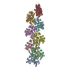
|
|---|---|
| 1 |
|
-要素
| #1: タンパク質 | / Alpha-actin-1 分子量: 41560.266 Da / 分子数: 8 / 由来タイプ: 天然 / 由来: (天然) Oryctolagus cuniculus (ウサギ) / 組織: Skeletal Muscle骨格筋 / 参照: UniProt: P68135#2: 化合物 | ChemComp-MG /   分子量: 24.305 Da / 分子数: 8 / 由来タイプ: 合成 / 式: Mg 分子量: 24.305 Da / 分子数: 8 / 由来タイプ: 合成 / 式: Mg#3: 化合物 | ChemComp-ADP / アデノシン二リン酸  分子量: 427.201 Da / 分子数: 8 / 由来タイプ: 合成 / 式: C10H15N5O10P2 / コメント: ADP, エネルギー貯蔵分子*YM 分子量: 427.201 Da / 分子数: 8 / 由来タイプ: 合成 / 式: C10H15N5O10P2 / コメント: ADP, エネルギー貯蔵分子*YM |
|---|
-実験情報
-実験
| 実験 | 手法: 電子顕微鏡法 |
|---|---|
| EM実験 | 試料の集合状態: FILAMENT / 3次元再構成法: らせん対称体再構成法 |
- 試料調製
試料調製
| 構成要素 | 名称: bare actin filament / タイプ: COMPLEX / 詳細: Actin filament in the ADP state / Entity ID: #1 / 由来: NATURAL | |||||||||||||||||||||||||||||||||||||||||||||
|---|---|---|---|---|---|---|---|---|---|---|---|---|---|---|---|---|---|---|---|---|---|---|---|---|---|---|---|---|---|---|---|---|---|---|---|---|---|---|---|---|---|---|---|---|---|---|
| 分子量 | 値: 0.042 MDa / 実験値: YES | |||||||||||||||||||||||||||||||||||||||||||||
| 由来(天然) | 生物種: Oryctolagus cuniculus (ウサギ) | |||||||||||||||||||||||||||||||||||||||||||||
| 緩衝液 | pH: 7.5 詳細: Buffer was filtered through 0.44 um filter and degassed. | |||||||||||||||||||||||||||||||||||||||||||||
| 緩衝液成分 |
| |||||||||||||||||||||||||||||||||||||||||||||
| 試料 | 濃度: 0.025 mg/ml / 包埋: NO / シャドウイング: NO / 染色: NO / 凍結: YES / 詳細: Filamentous bare actin | |||||||||||||||||||||||||||||||||||||||||||||
| 試料支持 | グリッドの材料: COPPER / グリッドのサイズ: 200 divisions/in. / グリッドのタイプ: C-flat-1.2/1.3 | |||||||||||||||||||||||||||||||||||||||||||||
| 急速凍結 | 装置: LEICA EM GP / 凍結剤: ETHANE / 湿度: 95 % / 凍結前の試料温度: 298 K 詳細: Sample was applied to a glow-discharged holey carbon grid, incubated for 60 seconds and blotted for 3 seconds from the backside with filter paper. |
- 電子顕微鏡撮影
| 顕微鏡 | モデル: FEI TECNAI 20 |
|---|---|
| 電子銃 | 電子線源: FIELD EMISSION GUN / 加速電圧: 200 kV / 照射モード: FLOOD BEAM |
| 電子レンズ | モード: BRIGHT FIELDBright-field microscopy / 倍率(公称値): 29000 X / 最大 デフォーカス(公称値): 3000 nm / 最小 デフォーカス(公称値): 1500 nm / Cs: 2 mm / C2レンズ絞り径: 100 µm / アライメント法: COMA FREE |
| 試料ホルダ | 凍結剤: NITROGEN 試料ホルダーモデル: GATAN 626 SINGLE TILT LIQUID NITROGEN CRYO TRANSFER HOLDER |
| 撮影 | 平均露光時間: 0.25 sec. / 電子線照射量: 1.5 e/Å2 / 検出モード: COUNTING フィルム・検出器のモデル: GATAN K2 SUMMIT (4k x 4k) 撮影したグリッド数: 1 / 実像数: 442 |
| 画像スキャン | サンプリングサイズ: 5 µm / 横: 3838 / 縦: 3710 / 動画フレーム数/画像: 24 / 利用したフレーム数/画像: 1-24 |
- 解析
解析
| EMソフトウェア |
| ||||||||||||||||||||||||||||||||||||||||||||||||||||
|---|---|---|---|---|---|---|---|---|---|---|---|---|---|---|---|---|---|---|---|---|---|---|---|---|---|---|---|---|---|---|---|---|---|---|---|---|---|---|---|---|---|---|---|---|---|---|---|---|---|---|---|---|---|
| CTF補正 | タイプ: PHASE FLIPPING AND AMPLITUDE CORRECTION | ||||||||||||||||||||||||||||||||||||||||||||||||||||
| らせん対称 | 回転角度/サブユニット: -166.6 ° / 軸方向距離/サブユニット: 28.11 Å / らせん対称軸の対称性: C1 | ||||||||||||||||||||||||||||||||||||||||||||||||||||
| 3次元再構成 | 解像度: 5.5 Å / 解像度の算出法: FSC 0.143 CUT-OFF / 粒子像の数: 63139 / アルゴリズム: FOURIER SPACE / クラス平均像の数: 1 / 対称性のタイプ: HELICAL | ||||||||||||||||||||||||||||||||||||||||||||||||||||
| 原子モデル構築 | B value: 350 / プロトコル: FLEXIBLE FIT / 空間: REAL 詳細: Initial models were assembled from 8 actins (3J8A) through rigid body docking in Chimera, followed by flexible fitting with DireX. Resulting models were subjected to MDFF. |
