 ムービー
ムービー コントローラー


+ データを開く

- 基本情報
基本情報
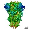
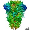
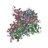
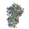
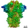
| 登録情報 | データベース: PDB / ID: 6bfu | |||||||||
|---|---|---|---|---|---|---|---|---|---|---|
| タイトル | Glycan shield and fusion activation of a deltacoronavirus spike glycoprotein fine-tuned for enteric infections | |||||||||
 要素 要素 | Spike protein スパイクタンパク質 スパイクタンパク質 | |||||||||
 キーワード キーワード | VIRAL PROTEIN (ウイルスタンパク質) / coronavirus spike glycoprotein / porcine deltacoronavirus / viral fusion protein / glycan shield / Structural Genomics (構造ゲノミクス) / Seattle Structural Genomics Center for Infectious Disease / SSGCID | |||||||||
| 機能・相同性 |  機能・相同性情報 機能・相同性情報: / endocytosis involved in viral entry into host cell / receptor-mediated virion attachment to host cell / membrane => GO:0016020 / fusion of virus membrane with host plasma membrane / fusion of virus membrane with host endosome membrane / エンベロープ (ウイルス) / virion membrane類似検索 - 分子機能 | |||||||||
| 生物種 |  Porcine deltacoronavirus (ウイルス) Porcine deltacoronavirus (ウイルス) | |||||||||
| 手法 | 電子顕微鏡法 / 単粒子再構成法 / クライオ電子顕微鏡法 / 解像度: 3.5 Å | |||||||||
 データ登録者 データ登録者 | Xiong, X. / Tortorici, M.A. / Snijder, S. / Yoshioka, C. / Walls, A.C. / Li, W. / Seattle Structural Genomics Center for Infectious Disease (SSGCID) / McGuire, A.T. / Rey, F.A. / Bosch, B.J. / Veesler, D. | |||||||||
| 資金援助 |  米国, 1件 米国, 1件
| |||||||||
 引用 引用 | ジャーナル: J Virol / 年: 2018 タイトル: Glycan Shield and Fusion Activation of a Deltacoronavirus Spike Glycoprotein Fine-Tuned for Enteric Infections. 著者: Xiaoli Xiong / M Alejandra Tortorici / Joost Snijder / Craig Yoshioka / Alexandra C Walls / Wentao Li / Andrew T McGuire / Félix A Rey / Berend-Jan Bosch / David Veesler /   要旨: Coronaviruses recently emerged as major human pathogens causing outbreaks of severe acute respiratory syndrome and Middle East respiratory syndrome. They utilize the spike (S) glycoprotein anchored ...Coronaviruses recently emerged as major human pathogens causing outbreaks of severe acute respiratory syndrome and Middle East respiratory syndrome. They utilize the spike (S) glycoprotein anchored in the viral envelope to mediate host attachment and fusion of the viral and cellular membranes to initiate infection. The S protein is a major determinant of the zoonotic potential of coronaviruses and is also the main target of the host humoral immune response. We report here the 3.5-Å-resolution cryo-electron microscopy structure of the S glycoprotein trimer from the pathogenic porcine deltacoronavirus (PDCoV), which belongs to the recently identified genus. Structural and glycoproteomics data indicate that the glycans of PDCoV S are topologically conserved compared with the human respiratory coronavirus NL63 S, resulting in similar surface areas being shielded from neutralizing antibodies and implying that both viruses are under comparable immune pressure in their respective hosts. The structure further reveals a shortened S' activation loop, containing a reduced number of basic amino acids, which participates in rendering the spike largely protease resistant. This property distinguishes PDCoV S from recently characterized betacoronavirus S proteins and suggests that the S protein of enterotropic PDCoV has evolved to tolerate the protease-rich environment of the small intestine and to fine-tune its fusion activation to avoid premature triggering and reduction of infectivity. Coronaviruses use transmembrane S glycoprotein trimers to promote host attachment and fusion of the viral and cellular membranes. We determined a near-atomic-resolution cryo-electron microscopy structure of the S ectodomain trimer from the pathogenic PDCoV, which is responsible for diarrhea in piglets and has had devastating consequences for the swine industry worldwide. Structural and glycoproteomics data reveal that PDCoV S is decorated with 78 N-linked glycans obstructing the protein surface to limit accessibility to neutralizing antibodies in a way reminiscent of what has recently been described for a human respiratory coronavirus. PDCoV S is largely protease resistant, which distinguishes it from most other characterized coronavirus S glycoproteins and suggests that enteric coronaviruses have evolved to fine-tune fusion activation in the protease-rich environment of the small intestine of infected hosts. | |||||||||
| 履歴 |
|
- 構造の表示
構造の表示
| ムービー |
ムービービューア |
|---|---|
| 構造ビューア | 分子: MolmilJmol/JSmol |
- ダウンロードとリンク
-ダウンロード
| PDBx/mmCIF形式 | 6bfu.cif.gz | 600 KB | 表示 | PDBx/mmCIF形式 |
|---|---|---|---|---|
| PDB形式 | pdb6bfu.ent.gz | 503.9 KB | 表示 | PDB形式 |
| PDBx/mmJSON形式 | 6bfu.json.gz | ツリー表示 | PDBx/mmJSON形式 | |
| その他 |  その他のダウンロード その他のダウンロード |
-検証レポート
| アーカイブディレクトリ | https://data.pdbj.org/pub/pdb/validation_reports/bf/6bfuftp://data.pdbj.org/pub/pdb/validation_reports/bf/6bfu | HTTPS FTP |
|---|
-関連構造データ


-リンク
 PDBj
PDBj- 集合体
集合体
| 登録構造単位 | 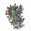
|
|---|---|
| 1 |
|
-要素
-タンパク質 , 1種, 3分子 ABC
| #1: タンパク質 | スパイクタンパク質 / spike glycoprotein 分子量: 112514.773 Da / 分子数: 3 / 由来タイプ: 組換発現 由来: (組換発現) Porcine deltacoronavirus (ウイルス)発現宿主:  Drosophila melanogaster (キイロショウジョウバエ) Drosophila melanogaster (キイロショウジョウバエ)参照: UniProt: A0A140ES81, UniProt: A0A140ESF1*PLUS |
|---|
-糖 , 5種, 63分子 
| #2: 多糖 | alpha-D-mannopyranose-(1-2)-alpha-D-mannopyranose-(1-3)-[alpha-D-mannopyranose-(1-6)]beta-D- ...alpha-D-mannopyranose-(1-2)-alpha-D-mannopyranose-(1-3)-[alpha-D-mannopyranose-(1-6)]beta-D-mannopyranose-(1-4)-2-acetamido-2-deoxy-beta-D-glucopyranose-(1-4)-2-acetamido-2-deoxy-beta-D-glucopyranose オリゴ糖 / 分子量: 1072.964 Da / 分子数: 9 / 由来タイプ: 組換発現#3: 多糖 | beta-D-mannopyranose-(1-4)-2-acetamido-2-deoxy-beta-D-glucopyranose-(1-4)-2-acetamido-2-deoxy-beta- ...beta-D-mannopyranose-(1-4)-2-acetamido-2-deoxy-beta-D-glucopyranose-(1-4)-2-acetamido-2-deoxy-beta-D-glucopyranose オリゴ糖 / 分子量: 586.542 Da / 分子数: 21 / 由来タイプ: 組換発現#4: 多糖 | 2-acetamido-2-deoxy-beta-D-glucopyranose-(1-4)-2-acetamido-2-deoxy-beta-D-glucopyranose オリゴ糖 / 分子量: 424.401 Da / 分子数: 6 / 由来タイプ: 組換発現#5: 多糖 | オリゴ糖 / 分子量: 910.823 Da / 分子数: 3 / 由来タイプ: 組換発現#6: 糖 | ChemComp-NAG / N-アセチルグルコサミン タイプ: D-saccharide, beta linking / 分子量: 221.208 Da / 分子数: 24 / 由来タイプ: 組換発現 / 式: C8H15NO6 タイプ: D-saccharide, beta linking / 分子量: 221.208 Da / 分子数: 24 / 由来タイプ: 組換発現 / 式: C8H15NO6 |
|---|
-実験情報
-実験
| 実験 | 手法: 電子顕微鏡法 |
|---|---|
| EM実験 | 試料の集合状態: PARTICLE / 3次元再構成法: 単粒子再構成法 |
- 試料調製
試料調製
| 構成要素 | 名称: Porcine deltacoronavirus spike glycoprotein / タイプ: COMPLEX / Entity ID: #1 / 由来: RECOMBINANT |
|---|---|
| 分子量 | 実験値: NO |
| 由来(天然) | 生物種: Porcine deltacoronavirus (ウイルス) |
| 由来(組換発現) | 生物種: Drosophila melanogaster (キイロショウジョウバエ) |
| 緩衝液 | pH: 8 |
| 試料 | 濃度: 0.5 mg/ml / 包埋: NO / シャドウイング: NO / 染色: NO / 凍結: YES |
| 急速凍結 | 凍結剤: ETHANE |
- 電子顕微鏡撮影
| 実験機器 |  モデル: Titan Krios / 画像提供: FEI Company |
|---|---|
| 顕微鏡 | モデル: FEI TITAN KRIOS |
| 電子銃 | 電子線源: FIELD EMISSION GUN / 加速電圧: 300 kV / 照射モード: FLOOD BEAM |
| 電子レンズ | モード: BRIGHT FIELDBright-field microscopy |
| 撮影 | 電子線照射量: 40 e/Å2 フィルム・検出器のモデル: GATAN K2 SUMMIT (4k x 4k) |
- 解析
解析
| EMソフトウェア |
| |||||||||
|---|---|---|---|---|---|---|---|---|---|---|
| CTF補正 | タイプ: PHASE FLIPPING AND AMPLITUDE CORRECTION | |||||||||
| 対称性 | 点対称性: C3 (3回回転対称) | |||||||||
| 3次元再構成 | 解像度: 3.5 Å / 解像度の算出法: FSC 0.143 CUT-OFF / 粒子像の数: 455710 / 対称性のタイプ: POINT |
