 ムービー
ムービー コントローラー


+ データを開く

- 基本情報
基本情報
| 登録情報 | データベース: PDB / ID: 6b3o | ||||||
|---|---|---|---|---|---|---|---|
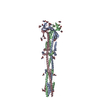
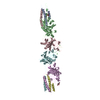
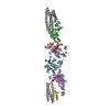
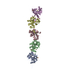
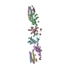
| タイトル | Tectonic conformational changes of a coronavirus spike glycoprotein promote membrane fusion | ||||||
 要素 要素 | Spike glycoprotein スパイクタンパク質 スパイクタンパク質 | ||||||
 キーワード キーワード | VIRAL PROTEIN (ウイルスタンパク質) / Coronavirus (オルトコロナウイルス亜科) / membrane fusion / MHV / SARS (重症急性呼吸器症候群) / MERS | ||||||
| 機能・相同性 |  機能・相同性情報 機能・相同性情報endocytosis involved in viral entry into host cell / host cell Golgi apparatus / host cell endoplasmic reticulum-Golgi intermediate compartment membrane / receptor-mediated virion attachment to host cell / fusion of virus membrane with host plasma membrane / fusion of virus membrane with host endosome membrane / エンベロープ (ウイルス) / host cell plasma membrane / virion membrane / 生体膜 / identical protein binding類似検索 - 分子機能 | ||||||
| 生物種 |  Murine coronavirus (マウス肝炎ウイルス) Murine coronavirus (マウス肝炎ウイルス) | ||||||
| 手法 | 電子顕微鏡法 / 単粒子再構成法 / クライオ電子顕微鏡法 / 解像度: 4.1 Å | ||||||
 データ登録者 データ登録者 | Walls, A.C. / Tortorici, M.A. / Snijder, J. / Xiong, X. / Bosch, B.J. / Rey, F.A. / Veesler, D. | ||||||
| 資金援助 |  米国, 1件 米国, 1件
| ||||||
 引用 引用 | ジャーナル: Proc Natl Acad Sci U S A / 年: 2017 タイトル: Tectonic conformational changes of a coronavirus spike glycoprotein promote membrane fusion. 著者: Alexandra C Walls / M Alejandra Tortorici / Joost Snijder / Xiaoli Xiong / Berend-Jan Bosch / Felix A Rey / David Veesler /   要旨: The tremendous pandemic potential of coronaviruses was demonstrated twice in the past few decades by two global outbreaks of deadly pneumonia. The coronavirus spike (S) glycoprotein initiates ...The tremendous pandemic potential of coronaviruses was demonstrated twice in the past few decades by two global outbreaks of deadly pneumonia. The coronavirus spike (S) glycoprotein initiates infection by promoting fusion of the viral and cellular membranes through conformational changes that remain largely uncharacterized. Here we report the cryoEM structure of a coronavirus S glycoprotein in the postfusion state, showing large-scale secondary, tertiary, and quaternary rearrangements compared with the prefusion trimer and rationalizing the free-energy landscape of this conformational machine. We also biochemically characterized the molecular events associated with refolding of the metastable prefusion S glycoprotein to the postfusion conformation using limited proteolysis, mass spectrometry, and single-particle EM. The observed similarity between postfusion coronavirus S and paramyxovirus F structures demonstrates that a conserved refolding trajectory mediates entry of these viruses and supports the evolutionary relatedness of their fusion subunits. Finally, our data provide a structural framework for understanding the mode of neutralization of antibodies targeting the fusion machinery and for engineering next-generation subunit vaccines or inhibitors against this medically important virus family. | ||||||
| 履歴 |
|
- 構造の表示
構造の表示
| ムービー |
ムービービューア |
|---|---|
| 構造ビューア | 分子: MolmilJmol/JSmol |
- ダウンロードとリンク
-ダウンロード
| PDBx/mmCIF形式 | 6b3o.cif.gz | 211.3 KB | 表示 | PDBx/mmCIF形式 |
|---|---|---|---|---|
| PDB形式 | pdb6b3o.ent.gz | 170.4 KB | 表示 | PDB形式 |
| PDBx/mmJSON形式 | 6b3o.json.gz | ツリー表示 | PDBx/mmJSON形式 | |
| その他 |  その他のダウンロード その他のダウンロード |
-検証レポート
| アーカイブディレクトリ | https://data.pdbj.org/pub/pdb/validation_reports/b3/6b3oftp://data.pdbj.org/pub/pdb/validation_reports/b3/6b3o | HTTPS FTP |
|---|
-関連構造データ


-リンク
 PDBj
PDBj- 集合体
集合体
| 登録構造単位 | 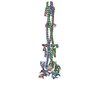
|
|---|---|
| 1 |
|
-要素
| #1: タンパク質 | スパイクタンパク質 / S glycoprotein / E2 / Peplomer protein 分子量: 66199.094 Da / 分子数: 3 / 断片: residues 718-1252 / 由来タイプ: 組換発現 由来: (組換発現) Murine coronavirus (マウス肝炎ウイルス)株: A59 / 遺伝子: S, 3 発現宿主:  Drosophila melanogaster (キイロショウジョウバエ) Drosophila melanogaster (キイロショウジョウバエ)参照: UniProt: P11224 配列の詳細 | The authors state that all the differences between the sequence provided and the sequence database ...The authors state that all the differences between the sequence provided and the sequence database reference are accounted for by modifications that they made to their construct: residues 1253-1254: BiP secretion signal, residues 1253-1254: linker, residues 1255-1284: GCN4 trimerization motif, residues 1285-1290: Thrombin cleavage site, residues 1291-1300: Strep-tag | |
|---|
-実験情報
-実験
| 実験 | 手法: 電子顕微鏡法 |
|---|---|
| EM実験 | 試料の集合状態: PARTICLE / 3次元再構成法: 単粒子再構成法 |
- 試料調製
試料調製
| 構成要素 | 名称: Mouse hepatitis virus spike glycoprotein (S2 subunit) in the postfusion conformation タイプ: COMPLEX / Entity ID: all / 由来: RECOMBINANT |
|---|---|
| 分子量 | 値: 0.18 MDa / 実験値: NO |
| 由来(天然) | 生物種: Murine hepatitis virus (マウス肝炎ウイルス) |
| 由来(組換発現) | 生物種: Drosophila melanogaster (キイロショウジョウバエ) |
| 緩衝液 | pH: 7.5 |
| 試料 | 包埋: NO / シャドウイング: NO / 染色: NO / 凍結: YES |
| 試料支持 | グリッドの材料: COPPER / グリッドのサイズ: 400 divisions/in. / グリッドのタイプ: C-flat |
| 急速凍結 | 凍結剤: ETHANE |
- 電子顕微鏡撮影
| 実験機器 |  モデル: Titan Krios / 画像提供: FEI Company |
|---|---|
| 顕微鏡 | モデル: FEI TITAN KRIOS |
| 電子銃 | 電子線源: FIELD EMISSION GUN / 加速電圧: 300 kV / 照射モード: FLOOD BEAM |
| 電子レンズ | モード: BRIGHT FIELDBright-field microscopy |
| 撮影 | 電子線照射量: 60 e/Å2 / 検出モード: COUNTING フィルム・検出器のモデル: GATAN K2 SUMMIT (4k x 4k) |
- 解析
解析
| EMソフトウェア |
| ||||||||||||||||||||||||||||||||
|---|---|---|---|---|---|---|---|---|---|---|---|---|---|---|---|---|---|---|---|---|---|---|---|---|---|---|---|---|---|---|---|---|---|
| CTF補正 | タイプ: PHASE FLIPPING AND AMPLITUDE CORRECTION | ||||||||||||||||||||||||||||||||
| 対称性 | 点対称性: C3 (3回回転対称) | ||||||||||||||||||||||||||||||||
| 3次元再構成 | 解像度: 4.1 Å / 解像度の算出法: FSC 0.143 CUT-OFF / 粒子像の数: 106000 / 対称性のタイプ: POINT | ||||||||||||||||||||||||||||||||
| 原子モデル構築 | プロトコル: AB INITIO MODEL / 空間: REAL |
