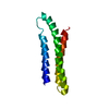
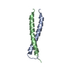
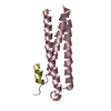
ジャーナル: Nat Struct Mol Biol / 年: 2017 タイトル: Cotranslational folding of spectrin domains via partially structured states. 著者: Ola B Nilsson / Adrian A Nickson / Jeffrey J Hollins / Stephan Wickles / Annette Steward / Roland Beckmann / Gunnar von Heijne / Jane Clarke / 要旨: How do the key features of protein folding, elucidated from studies on native, isolated proteins, manifest in cotranslational folding on the ribosome? Using a well-characterized family of homologous ...How do the key features of protein folding, elucidated from studies on native, isolated proteins, manifest in cotranslational folding on the ribosome? Using a well-characterized family of homologous α-helical proteins with a range of biophysical properties, we show that spectrin domains can fold vectorially on the ribosome and may do so via a pathway different from that of the isolated domain. We use cryo-EM to reveal a folded or partially folded structure, formed in the vestibule of the ribosome. Our results reveal that it is not possible to predict which domains will fold within the ribosome on the basis of the folding behavior of isolated domains; instead, we propose that a complex balance of the rate of folding, the rate of translation and the lifetime of folded or partly folded states will determine whether folding occurs cotranslationally on actively translating ribosomes.
電子線照射量: 2.4 e/Å2 フィルム・検出器のモデル: FEI FALCON II (4k x 4k)
-
解析
EMソフトウェア
ID
名称
バージョン
カテゴリ
2
EM-Tools
画像取得
4
CTFFIND
4
CTF補正
9
SPIDER
初期オイラー角割当
10
SPIDER
最終オイラー角割当
11
SPIDER
分類
12
SPIDER
3次元再構成
CTF補正
タイプ: NONE
3次元再構成
解像度: 4.8 Å / 解像度の算出法: FSC 0.143 CUT-OFF / 粒子像の数: 46067 詳細: To exclude potential overfitting, the data were processed using a frequency limited refinement protocol by truncating high frequencies (low-pass filter at 8 A) 対称性のタイプ: POINT
 ムービー
ムービー コントローラー


 データを開く
 基本情報
基本情報 要素
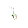
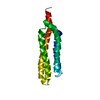
要素 スペクトリン
  キーワード
キーワード 機能・相同性情報
 データ登録者
 引用
引用


 構造の表示
構造の表示 ダウンロードとリンク
 その他のダウンロード
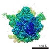


 PDBj
PDBj
 集合体
集合体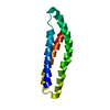
 試料調製
試料調製 電子顕微鏡撮影
 解析
解析