 ムービー
ムービー コントローラー


+ データを開く

- 基本情報
基本情報
| 登録情報 | データベース: PDB / ID: 5h1s | ||||||
|---|---|---|---|---|---|---|---|
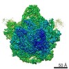
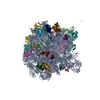
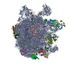
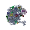
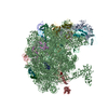
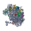
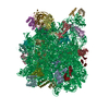
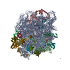
| タイトル | Structure of the large subunit of the chloro-ribosome | ||||||
 要素 要素 |
| ||||||
 キーワード キーワード |  RIBOSOME (リボソーム) / Cryo-EM (低温電子顕微鏡法) / chloro-ribosome RIBOSOME (リボソーム) / Cryo-EM (低温電子顕微鏡法) / chloro-ribosome | ||||||
| 機能・相同性 |  機能・相同性情報 機能・相同性情報plastid translation / mitochondrial large ribosomal subunit / mitochondrial translation / 葉緑体 / DNA-templated transcription termination / large ribosomal subunit rRNA binding / large ribosomal subunit / cytoplasmic translation / 5S rRNA binding / cytosolic large ribosomal subunit ...plastid translation / mitochondrial large ribosomal subunit / mitochondrial translation / 葉緑体 / DNA-templated transcription termination / large ribosomal subunit rRNA binding / large ribosomal subunit / cytoplasmic translation / 5S rRNA binding / cytosolic large ribosomal subunit / transferase activity / negative regulation of translation / rRNA binding / リボソーム / structural constituent of ribosome / ribonucleoprotein complex / 翻訳 (生物学) / mRNA binding / ミトコンドリア / RNA binding類似検索 - 分子機能 | ||||||
| 生物種 |  Spinacia oleracea (ホウレンソウ) Spinacia oleracea (ホウレンソウ) | ||||||
| 手法 | 電子顕微鏡法 / 単粒子再構成法 / クライオ電子顕微鏡法 / 解像度: 3.5 Å | ||||||
 データ登録者 データ登録者 | Ahmed, T. / Yin, Z. / Bhushan, S. | ||||||
| 資金援助 |  シンガポール, 1件 シンガポール, 1件
| ||||||
 引用 引用 | ジャーナル: Sci Rep / 年: 2016 タイトル: Cryo-EM structure of the large subunit of the spinach chloroplast ribosome. 著者: Tofayel Ahmed / Zhan Yin / Shashi Bhushan / 要旨: Protein synthesis in the chloroplast is mediated by the chloroplast ribosome (chloro-ribosome). Overall architecture of the chloro-ribosome is considerably similar to the Escherichia coli (E. coli) ...Protein synthesis in the chloroplast is mediated by the chloroplast ribosome (chloro-ribosome). Overall architecture of the chloro-ribosome is considerably similar to the Escherichia coli (E. coli) ribosome but certain differences are evident. The chloro-ribosome proteins are generally larger because of the presence of chloroplast-specific extensions in their N- and C-termini. The chloro-ribosome harbours six plastid-specific ribosomal proteins (PSRPs); four in the small subunit and two in the large subunit. Deletions and insertions occur throughout the rRNA sequence of the chloro-ribosome (except for the conserved peptidyl transferase center region) but the overall length of the rRNAs do not change significantly, compared to the E. coli. Although, recent advancements in cryo-electron microscopy (cryo-EM) have provided detailed high-resolution structures of ribosomes from many different sources, a high-resolution structure of the chloro-ribosome is still lacking. Here, we present a cryo-EM structure of the large subunit of the chloro-ribosome from spinach (Spinacia oleracea) at an average resolution of 3.5 Å. High-resolution map enabled us to localize and model chloro-ribosome proteins, chloroplast-specific protein extensions, two PSRPs (PSRP5 and 6) and three rRNA molecules present in the chloro-ribosome. Although comparable to E. coli, the polypeptide tunnel and the tunnel exit site show chloroplast-specific features. | ||||||
| 履歴 |
|
- 構造の表示
構造の表示
| ムービー |
ムービービューア |
|---|---|
| 構造ビューア | 分子: MolmilJmol/JSmol |
- ダウンロードとリンク
-ダウンロード
| PDBx/mmCIF形式 | 5h1s.cif.gz | 2 MB | 表示 | PDBx/mmCIF形式 |
|---|---|---|---|---|
| PDB形式 | pdb5h1s.ent.gz | 1.5 MB | 表示 | PDB形式 |
| PDBx/mmJSON形式 | 5h1s.json.gz | ツリー表示 | PDBx/mmJSON形式 | |
| その他 |  その他のダウンロード その他のダウンロード |
-検証レポート
| アーカイブディレクトリ | https://data.pdbj.org/pub/pdb/validation_reports/h1/5h1sftp://data.pdbj.org/pub/pdb/validation_reports/h1/5h1s | HTTPS FTP |
|---|
-関連構造データ


-リンク
 PDBj
PDBj


- 集合体
集合体
| 登録構造単位 | 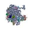
|
|---|---|
| 1 |
|
-要素
-RNA鎖 , 3種, 3分子 ACB
| #1: RNA鎖 | 23SリボソームRNA 分子量: 911344.250 Da / 分子数: 1 / 由来タイプ: 天然 / 由来: (天然) Spinacia oleracea (ホウレンソウ) / 参照: GenBank: AJ400848 |
|---|---|
| #2: RNA鎖 | 分子量: 34334.488 Da / 分子数: 1 / 由来タイプ: 天然 / 由来: (天然) Spinacia oleracea (ホウレンソウ) / 参照: GenBank: 175996 |
| #3: RNA鎖 | 5SリボソームRNA 分子量: 39014.184 Da / 分子数: 1 / 由来タイプ: 天然 / 由来: (天然) Spinacia oleracea (ホウレンソウ) |
+50S ribosomal protein ... , 29種, 29分子 LMNOPQRSTUVWXYZEbcdefFGHIJgah
-実験情報
-実験
| 実験 | 手法: 電子顕微鏡法 |
|---|---|
| EM実験 | 試料の集合状態: PARTICLE / 3次元再構成法: 単粒子再構成法 |
- 試料調製
試料調製
| 構成要素 | 名称: Chloro-ribosome 50S / タイプ: RIBOSOME / Entity ID: all / 由来: NATURAL |
|---|---|
| 分子量 | 値: 1.7 MDa / 実験値: NO |
| 由来(天然) | 生物種: Spinacia oleracea (ホウレンソウ) |
| 緩衝液 | pH: 7.6 詳細: 20 mM Tris HCl, pH 7.6, 100 mM KCl, 10 mM MgOAc, 100 mM sucrose, 7 mM 2-mercaptoethanol, 1 unit/ml RNase inhibitor, 0.1% protease inhibitor |
| 試料 | 包埋: NO / シャドウイング: NO / 染色: NO / 凍結: YES |
| 試料支持 | グリッドの材料: COPPER / グリッドのサイズ: 300 divisions/in. / グリッドのタイプ: Quantifoil |
| 急速凍結 | 装置: FEI VITROBOT MARK IV / 凍結剤: ETHANE / 湿度: 100 % / 凍結前の試料温度: 277 K |
- 電子顕微鏡撮影
| 実験機器 |  モデル: Talos Arctica / 画像提供: FEI Company |
|---|---|
| 顕微鏡 | モデル: FEI TECNAI ARCTICA |
| 電子銃 | 電子線源: FIELD EMISSION GUN / 加速電圧: 200 kV / 照射モード: FLOOD BEAM |
| 電子レンズ | モード: BRIGHT FIELDBright-field microscopy / 倍率(公称値): 78000 X / 倍率(補正後): 109375 X / 最大 デフォーカス(公称値): 2500 nm / 最小 デフォーカス(公称値): 200 nm / Calibrated defocus min: 200 nm / 最大 デフォーカス(補正後): 2500 nm / Cs: 2.7 mm / C2レンズ絞り径: 100 µm / アライメント法: COMA FREE |
| 試料ホルダ | 凍結剤: NITROGEN 試料ホルダーモデル: FEI TITAN KRIOS AUTOGRID HOLDER |
| 撮影 | 電子線照射量: 26 e/Å2 / 検出モード: INTEGRATING フィルム・検出器のモデル: FEI FALCON II (4k x 4k) 撮影したグリッド数: 1 / 実像数: 1590 |
| 画像スキャン | サンプリングサイズ: 14 µm / 横: 4096 / 縦: 4096 / 動画フレーム数/画像: 7 / 利用したフレーム数/画像: 1-7 |
- 解析
解析
| ソフトウェア | 名称: PHENIX / バージョン: dev_2210: / 分類: 精密化 | ||||||||||||||||||||||||
|---|---|---|---|---|---|---|---|---|---|---|---|---|---|---|---|---|---|---|---|---|---|---|---|---|---|
| EMソフトウェア |
| ||||||||||||||||||||||||
| CTF補正 | タイプ: PHASE FLIPPING ONLY | ||||||||||||||||||||||||
| 粒子像の選択 | 選択した粒子像数: 338305 | ||||||||||||||||||||||||
| 対称性 | 点対称性: C1 (非対称) | ||||||||||||||||||||||||
| 3次元再構成 | 解像度: 3.5 Å / 解像度の算出法: FSC 0.143 CUT-OFF / 粒子像の数: 174949 / アルゴリズム: FOURIER SPACE / クラス平均像の数: 20 / 対称性のタイプ: POINT | ||||||||||||||||||||||||
| 拘束条件 |
|
