 ムービー
ムービー コントローラー


+ データを開く

- 基本情報
基本情報
| 登録情報 | データベース: PDB / ID: 5000000000000 | ||||||
|---|---|---|---|---|---|---|---|
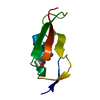
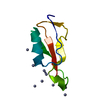
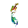
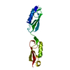
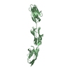
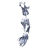
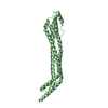
| タイトル | Crystal Structure of PASTA Domains 2, 3 and 4 of Mycobacterium tuberculosis Protein Kinase B | ||||||
 要素 要素 | Serine/threonine-protein kinase PknB | ||||||
 キーワード キーワード |  TRANSFERASE (転移酵素) / kinase (キナーゼ) / extracellular sensor domain / peptidoglycan binding (ペプチドグリカン) / Structural Genomics (構造ゲノミクス) / TB Structural Genomics Consortium / TBSGC TRANSFERASE (転移酵素) / kinase (キナーゼ) / extracellular sensor domain / peptidoglycan binding (ペプチドグリカン) / Structural Genomics (構造ゲノミクス) / TB Structural Genomics Consortium / TBSGC | ||||||
| 機能・相同性 |  機能・相同性情報 機能・相同性情報negative regulation of growth rate / acetyltransferase activator activity / negative regulation of catalytic activity / negative regulation of fatty acid biosynthetic process / response to host immune response / positive regulation of catalytic activity / positive regulation of DNA binding / peptidoglycan biosynthetic process / peptidoglycan-based cell wall / negative regulation of protein binding ...negative regulation of growth rate / acetyltransferase activator activity / negative regulation of catalytic activity / negative regulation of fatty acid biosynthetic process / response to host immune response / positive regulation of catalytic activity / positive regulation of DNA binding / peptidoglycan biosynthetic process / peptidoglycan-based cell wall / negative regulation of protein binding / manganese ion binding / regulation of cell shape / protein autophosphorylation / non-specific serine/threonine protein kinase / protein kinase activity / protein phosphorylation / protein serine kinase activity / protein serine/threonine kinase activity / ATP binding / identical protein binding / 細胞膜類似検索 - 分子機能 | ||||||
| 生物種 |   Mycobacterium tuberculosis (結核菌) Mycobacterium tuberculosis (結核菌) | ||||||
| 手法 | X線回折 / シンクロトロン / 分子置換 / 解像度: 2.208 Å | ||||||
 データ登録者 データ登録者 | Prigozhin, D.M. / TB Structural Genomics Consortium (TBSGC) | ||||||
| 資金援助 |  米国, 1件 米国, 1件
| ||||||
 引用 引用 | ジャーナル: J.Biol.Chem. / 年: 2016 タイトル: Structural and Genetic Analyses of the Mycobacterium tuberculosis Protein Kinase B Sensor Domain Identify a Potential Ligand-binding Site. 著者: Prigozhin, D.M. / Papavinasasundaram, K.G. / Baer, C.E. / Murphy, K.C. / Moskaleva, A. / Chen, T.Y. / Alber, T. / Sassetti, C.M. | ||||||
| 履歴 |
|
- 構造の表示
構造の表示
| 構造ビューア | 分子: MolmilJmol/JSmol |
|---|
- ダウンロードとリンク
-ダウンロード
| PDBx/mmCIF形式 | 5e12.cif.gz | 88.3 KB | 表示 | PDBx/mmCIF形式 |
|---|---|---|---|---|
| PDB形式 | pdb5e12.ent.gz | 65.9 KB | 表示 | PDB形式 |
| PDBx/mmJSON形式 | 5e12.json.gz | ツリー表示 | PDBx/mmJSON形式 | |
| その他 |  その他のダウンロード その他のダウンロード |
-検証レポート
| アーカイブディレクトリ | https://data.pdbj.org/pub/pdb/validation_reports/e1/5e12ftp://data.pdbj.org/pub/pdb/validation_reports/e1/5e12 | HTTPS FTP |
|---|
-関連構造データ


-リンク
 PDBj
PDBj


- 集合体
集合体
| 登録構造単位 | 
| ||||||||
|---|---|---|---|---|---|---|---|---|---|
| 1 | 
| ||||||||
| 2 | 
| ||||||||
| 単位格子 |
|
-要素
| #1: タンパク質 | 分子量: 21360.895 Da / 分子数: 2 / 断片: UNP residues 423-626 / 由来タイプ: 組換発現 由来: (組換発現) Mycobacterium tuberculosis (結核菌)株: ATCC 25618 / H37Rv / 遺伝子: pknB, Rv0014c, MTCY10H4.14c / 発現宿主: Escherichia coli (大腸菌)参照: UniProt: P9WI81, non-specific serine/threonine protein kinase#2: 化合物 | ChemComp-FLC / | クエン酸  分子量: 189.100 Da / 分子数: 1 / 由来タイプ: 合成 / 式: C6H5O7 分子量: 189.100 Da / 分子数: 1 / 由来タイプ: 合成 / 式: C6H5O7#3: 水 | ChemComp-HOH / | 水 分子量: 18.015 Da / 分子数: 141 / 由来タイプ: 天然 / 式: H2O 分子量: 18.015 Da / 分子数: 141 / 由来タイプ: 天然 / 式: H2O |
|---|
-実験情報
-実験
| 実験 | 手法: X線回折 / 使用した結晶の数: 1 |
|---|
- 試料調製
試料調製
| 結晶 | マシュー密度: 2.15 Å3/Da / 溶媒含有率: 42.75 % |
|---|---|
| 結晶化 | 温度: 291 K / 手法: 蒸気拡散法 / 詳細: 0.1 M citrate pH 3.5, 25% PEG 3350 |
-データ収集
| 回折 | 平均測定温度: 100 K | ||||||||||||||||||||||||||||||||||||||||||||||||||||||||||||||||||
|---|---|---|---|---|---|---|---|---|---|---|---|---|---|---|---|---|---|---|---|---|---|---|---|---|---|---|---|---|---|---|---|---|---|---|---|---|---|---|---|---|---|---|---|---|---|---|---|---|---|---|---|---|---|---|---|---|---|---|---|---|---|---|---|---|---|---|---|
| 放射光源 | 由来: シンクロトロン / サイト: ALS / ビームライン: 8.3.1 / 波長: 1.116 Å | ||||||||||||||||||||||||||||||||||||||||||||||||||||||||||||||||||
| 検出器 | タイプ: ADSC QUANTUM 315r / 検出器: CCD / 日付: 2011年2月19日 | ||||||||||||||||||||||||||||||||||||||||||||||||||||||||||||||||||
| 放射 | モノクロメーター: Double flat crystal, Si(111) / プロトコル: SINGLE WAVELENGTH / 単色(M)・ラウエ(L): M / 散乱光タイプ: x-ray | ||||||||||||||||||||||||||||||||||||||||||||||||||||||||||||||||||
| 放射波長 | 波長: 1.116 Å / 相対比: 1 | ||||||||||||||||||||||||||||||||||||||||||||||||||||||||||||||||||
| 反射 | 解像度: 2.2→41.902 Å / Num. obs: 18775 / % possible obs: 99.9 % / 冗長度: 4 % / Biso Wilson estimate: 25.98 Å2 / Rmerge(I) obs: 0.138 / Χ2: 1.154 / Net I/av σ(I): 7.79 / Net I/σ(I): 7.8 / Num. measured all: 74634 | ||||||||||||||||||||||||||||||||||||||||||||||||||||||||||||||||||
| 反射 シェル | Diffraction-ID: 1 / Rejects: 0
|
-位相決定
| 位相決定 | 手法: 分子置換 |
|---|
- 解析
解析
| ソフトウェア |
| ||||||||||||||||||||||||||||||||||||||||||||||||||||||||||||||||||||||||||||||||||||||||||||||||||
|---|---|---|---|---|---|---|---|---|---|---|---|---|---|---|---|---|---|---|---|---|---|---|---|---|---|---|---|---|---|---|---|---|---|---|---|---|---|---|---|---|---|---|---|---|---|---|---|---|---|---|---|---|---|---|---|---|---|---|---|---|---|---|---|---|---|---|---|---|---|---|---|---|---|---|---|---|---|---|---|---|---|---|---|---|---|---|---|---|---|---|---|---|---|---|---|---|---|---|---|
| 精密化 | 構造決定の手法: 分子置換 / 解像度: 2.208→41.902 Å / SU ML: 0.27 / 交差検証法: FREE R-VALUE / σ(F): 1.33 / 位相誤差: 27.4 / 立体化学のターゲット値: ML
| ||||||||||||||||||||||||||||||||||||||||||||||||||||||||||||||||||||||||||||||||||||||||||||||||||
| 溶媒の処理 | 減衰半径: 0.6 Å / VDWプローブ半径: 0.9 Å / 溶媒モデル: FLAT BULK SOLVENT MODEL | ||||||||||||||||||||||||||||||||||||||||||||||||||||||||||||||||||||||||||||||||||||||||||||||||||
| 原子変位パラメータ | Biso max: 80.21 Å2 / Biso mean: 30.8683 Å2 / Biso min: 7.53 Å2 | ||||||||||||||||||||||||||||||||||||||||||||||||||||||||||||||||||||||||||||||||||||||||||||||||||
| 精密化ステップ | サイクル: final / 解像度: 2.208→41.902 Å
| ||||||||||||||||||||||||||||||||||||||||||||||||||||||||||||||||||||||||||||||||||||||||||||||||||
| 拘束条件 |
| ||||||||||||||||||||||||||||||||||||||||||||||||||||||||||||||||||||||||||||||||||||||||||||||||||
| LS精密化 シェル | Refine-ID: X-RAY DIFFRACTION / Total num. of bins used: 13
|
