 ムービー
ムービー コントローラー


+ データを開く

- 基本情報
基本情報
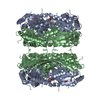
| 登録情報 | データベース: PDB / ID: 3m9i | ||||||
|---|---|---|---|---|---|---|---|
| タイトル | Electron crystallographic structure of lens Aquaporin-0 (AQP0) (lens MIP) in E. coli polar lipids | ||||||
 要素 要素 | Lens fiber major intrinsic protein | ||||||
 キーワード キーワード | MEMBRANE PROTEIN (膜タンパク質) / water channel (アクアポリン) / lens / lipid-protein interactions | ||||||
| 機能・相同性 |  機能・相同性情報 機能・相同性情報gap junction-mediated intercellular transport / water channel activity / water transport / structural constituent of eye lens / ギャップ結合 / response to stimulus / lens development in camera-type eye / positive regulation of cell adhesion / 視覚 / protein homotetramerization ...gap junction-mediated intercellular transport / water channel activity / water transport / structural constituent of eye lens / ギャップ結合 / response to stimulus / lens development in camera-type eye / positive regulation of cell adhesion / 視覚 / protein homotetramerization / calmodulin binding / 小胞体 / 細胞膜類似検索 - 分子機能 | ||||||
| 生物種 |  Ovis aries (ヒツジ) Ovis aries (ヒツジ) | ||||||
| 手法 | 電子線結晶学 / 分子置換 / クライオ電子顕微鏡法 / 解像度: 2.5 Å | ||||||
 データ登録者 データ登録者 | Hite, R.K. / Li, Z. / Walz, T. | ||||||
 引用 引用 | ジャーナル: EMBO J / 年: 2010 タイトル: Principles of membrane protein interactions with annular lipids deduced from aquaporin-0 2D crystals. 著者: Richard K Hite / Zongli Li / Thomas Walz /  要旨: We have previously described the interactions of aquaporin-0 (AQP0) with dimyristoyl phosphatidylcholine (DMPC) lipids. We have now determined the 2.5 A structure of AQP0 in two-dimensional (2D) ...We have previously described the interactions of aquaporin-0 (AQP0) with dimyristoyl phosphatidylcholine (DMPC) lipids. We have now determined the 2.5 A structure of AQP0 in two-dimensional (2D) crystals formed with Escherichia coli polar lipids (EPLs), which differ from DMPC both in headgroups and acyl chains. Comparison of the two structures shows that AQP0 does not adapt to the different length of the acyl chains in EPLs and that the distance between the phosphodiester groups in the two leaflets of the DMPC and EPL bilayers is almost identical. The EPL headgroups interact differently with AQP0 than do those of DMPC, but the acyl chains in the EPL and DMPC bilayers occupy similar positions. The interactions of annular lipids with membrane proteins seem to be driven by the propensity of the acyl chains to fill gaps in the protein surface. Interactions of the lipid headgroups may be responsible for the specific interactions found in tightly bound lipids but seem to have a negligible effect on interactions of generic annular lipids with membrane proteins. | ||||||
| 履歴 |
|
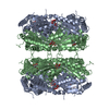
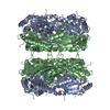
- 構造の表示
構造の表示
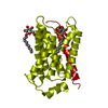
| ムービー |
ムービービューア |
|---|---|
| 構造ビューア | 分子: MolmilJmol/JSmol |
- ダウンロードとリンク
-ダウンロード
| PDBx/mmCIF形式 | 3m9i.cif.gz | 63.3 KB | 表示 | PDBx/mmCIF形式 |
|---|---|---|---|---|
| PDB形式 | pdb3m9i.ent.gz | 45.9 KB | 表示 | PDB形式 |
| PDBx/mmJSON形式 | 3m9i.json.gz | ツリー表示 | PDBx/mmJSON形式 | |
| その他 |  その他のダウンロード その他のダウンロード |
-検証レポート
| アーカイブディレクトリ | https://data.pdbj.org/pub/pdb/validation_reports/m9/3m9iftp://data.pdbj.org/pub/pdb/validation_reports/m9/3m9i | HTTPS FTP |
|---|
-関連構造データ
| 関連構造データ |  2b6oS S: 精密化の開始モデル |
|---|---|
| 類似構造データ |


-リンク
 PDBj
PDBj
- 集合体
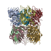
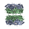
集合体
| 登録構造単位 | 
| ||||||||
|---|---|---|---|---|---|---|---|---|---|
| 1 | x 8
| ||||||||
| 単位格子 |
| ||||||||
| 詳細 | The octamer can be generated by applying P422 symmetry. |
-要素
| #1: タンパク質 | / Aquaporin-0 分子量: 23514.447 Da / 分子数: 1 / 断片: UNP residues 7 to 226 / 由来タイプ: 天然 / 由来: (天然) Ovis aries (ヒツジ) / 参照: UniProt: Q6J8I9 | ||
|---|---|---|---|
| #2: 化合物 | ChemComp-3PE / ホスファチジルエタノールアミン  分子量: 748.065 Da / 分子数: 7 / 由来タイプ: 合成 / 式: C41H82NO8P / コメント: リン脂質*YM 分子量: 748.065 Da / 分子数: 7 / 由来タイプ: 合成 / 式: C41H82NO8P / コメント: リン脂質*YM#3: 水 | ChemComp-HOH / | 水 分子量: 18.015 Da / 分子数: 8 / 由来タイプ: 天然 / 式: H2O 分子量: 18.015 Da / 分子数: 8 / 由来タイプ: 天然 / 式: H2O |
-実験情報
-実験
| 実験 | 手法: 電子線結晶学 / 使用した結晶の数: 281 |
|---|---|
| EM実験 | 試料の集合状態: 2D ARRAY / 3次元再構成法: 電子線結晶学 |
- 試料調製
試料調製
| 構成要素 | 名称: lens Aquaporin 0 / タイプ: COMPLEX |
|---|---|
| 緩衝液 | pH: 8 |
| 試料 | 包埋: NO / シャドウイング: NO / 染色: NO / 凍結: YES 詳細: Purified membranes were solubilized in 4% (w/v) octyl glucoside in 10 mM Tris (pH 8.0) for 30 min at 22C |
| 結晶化 | 温度: 300 K / 手法: microdialysis / pH: 6 詳細: 100 mM NaCl, 50mM MgCl2, 10mM MES pH 6.0, MICRODIALYSIS, temperature 300K |
-データ収集
| 実験機器 |  モデル: Tecnai Polara / 画像提供: FEI Company |
|---|---|
| 顕微鏡 | モデル: FEI POLARA 300 |
| 電子銃 | 電子線源: FIELD EMISSION GUN / 加速電圧: 300 kV / 照射モード: FLOOD BEAM |
| 電子レンズ | モード: DIFFRACTION回折 |
| 撮影 | フィルム・検出器のモデル: GENERIC GATAN (4k x 4k) |
| 回折 | 平均測定温度: 279 K |
| 放射光源 | 由来: ELECTRON MICROSCOPE / タイプ: その他 |
| 検出器 | タイプ: Gatan / 検出器: CCD / 日付: 2009年2月24日 |
| 反射 | 解像度: 2.5→15.89 Å / Num. all: 14417 / Num. obs: 14417 / % possible obs: 92.3 % / Observed criterion σ(F): 0 / Observed criterion σ(I): 0 / 冗長度: 8.1 % / Biso Wilson estimate: 14.9 Å2 / Rmerge(I) obs: 0.226 / Rsym value: 0.189 |
| 反射 シェル | 解像度: 2.5→2.66 Å / 冗長度: 4 % / Num. unique all: 1935 / % possible all: 83.7 |
- 解析
解析
| ソフトウェア |
| |||||||||||||||||||||||||
|---|---|---|---|---|---|---|---|---|---|---|---|---|---|---|---|---|---|---|---|---|---|---|---|---|---|---|
| 精密化 | 構造決定の手法: 分子置換 開始モデル: PDB entry 2B6O 解像度: 2.5→15.89 Å / Rfactor Rfree error: 0.007 / Data cutoff high absF: 2341911.44 / Data cutoff low absF: 0 / Isotropic thermal model: RESTRAINED / 交差検証法: THROUGHOUT / σ(F): 0 / 立体化学のターゲット値: Engh & Huber / 詳細: BULK SOLVENT MODEL USED
| |||||||||||||||||||||||||
| 溶媒の処理 | 溶媒モデル: FLAT MODEL / Bsol: 44.0682 Å2 / ksol: 0.1 e/Å3 | |||||||||||||||||||||||||
| 原子変位パラメータ | Biso mean: 48.6 Å2
| |||||||||||||||||||||||||
| Refine analyze |
| |||||||||||||||||||||||||
| 精密化ステップ | サイクル: LAST / 解像度: 2.5→15.89 Å
| |||||||||||||||||||||||||
| 拘束条件 |
| |||||||||||||||||||||||||
| Refine LS restraints NCS | NCS model details: NONE | |||||||||||||||||||||||||
| LS精密化 シェル | 解像度: 2.5→2.66 Å / Rfactor Rfree error: 0.029 / Total num. of bins used: 6
| |||||||||||||||||||||||||
| Xplor file |
|
