ムービー
ムービー コントローラー


+ データを開く

- 基本情報
基本情報
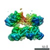
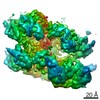
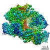
| 登録情報 | データベース: PDB / ID: 3j95 | ||||||
|---|---|---|---|---|---|---|---|
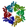
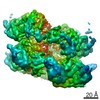
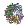
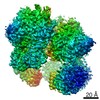
| タイトル | Structure of ADP-bound N-ethylmaleimide sensitive factor determined by single particle cryoelectron microscopy | ||||||
 要素 要素 | Vesicle-fusing ATPase | ||||||
 キーワード キーワード | HYDROLASE (加水分解酵素) / ATPases associated with diverse cellular activities | ||||||
| 機能・相同性 |  機能・相同性情報 機能・相同性情報SNARE complex disassembly / ATP-dependent protein disaggregase activity / vesicle-fusing ATPase / syntaxin-1 binding / positive regulation of receptor recycling / ionotropic glutamate receptor binding / SNARE binding / PDZ domain binding / intracellular protein transport / potassium ion transport ...SNARE complex disassembly / ATP-dependent protein disaggregase activity / vesicle-fusing ATPase / syntaxin-1 binding / positive regulation of receptor recycling / ionotropic glutamate receptor binding / SNARE binding / PDZ domain binding / intracellular protein transport / potassium ion transport / positive regulation of protein catabolic process / midbody / protein-containing complex binding / protein kinase binding / ゴルジ体 / ATP hydrolysis activity / ATP binding / identical protein binding / metal ion binding / 細胞膜 / 細胞質基質類似検索 - 分子機能 | ||||||
| 生物種 |   Cricetulus griseus (モンゴルキヌゲネズミ) Cricetulus griseus (モンゴルキヌゲネズミ) | ||||||
| 手法 | 電子顕微鏡法 / 単粒子再構成法 / クライオ電子顕微鏡法 / 解像度: 7.6 Å | ||||||
 データ登録者 データ登録者 | Zhao, M. / Wu, S. / Cheng, Y. / Brunger, A.T. | ||||||
 引用 引用 | ジャーナル: Nature / 年: 2015 タイトル: Mechanistic insights into the recycling machine of the SNARE complex. 著者: Minglei Zhao / Shenping Wu / Qiangjun Zhou / Sandro Vivona / Daniel J Cipriano / Yifan Cheng / Axel T Brunger /  要旨: Evolutionarily conserved SNARE (soluble N-ethylmaleimide sensitive factor attachment protein receptors) proteins form a complex that drives membrane fusion in eukaryotes. The ATPase NSF (N- ...Evolutionarily conserved SNARE (soluble N-ethylmaleimide sensitive factor attachment protein receptors) proteins form a complex that drives membrane fusion in eukaryotes. The ATPase NSF (N-ethylmaleimide sensitive factor), together with SNAPs (soluble NSF attachment protein), disassembles the SNARE complex into its protein components, making individual SNAREs available for subsequent rounds of fusion. Here we report structures of ATP- and ADP-bound NSF, and the NSF/SNAP/SNARE (20S) supercomplex determined by single-particle electron cryomicroscopy at near-atomic to sub-nanometre resolution without imposing symmetry. Large, potentially force-generating, conformational differences exist between ATP- and ADP-bound NSF. The 20S supercomplex exhibits broken symmetry, transitioning from six-fold symmetry of the NSF ATPase domains to pseudo four-fold symmetry of the SNARE complex. SNAPs interact with the SNARE complex with an opposite structural twist, suggesting an unwinding mechanism. The interfaces between NSF, SNAPs, and SNAREs exhibit characteristic electrostatic patterns, suggesting how one NSF/SNAP species can act on many different SNARE complexes. | ||||||
| 履歴 |
|
- 構造の表示
構造の表示
| ムービー |
ムービービューア |
|---|---|
| 構造ビューア | 分子: MolmilJmol/JSmol |
- ダウンロードとリンク
-ダウンロード
| PDBx/mmCIF形式 | 3j95.cif.gz | 483.6 KB | 表示 | PDBx/mmCIF形式 |
|---|---|---|---|---|
| PDB形式 | pdb3j95.ent.gz | 361.3 KB | 表示 | PDB形式 |
| PDBx/mmJSON形式 | 3j95.json.gz | ツリー表示 | PDBx/mmJSON形式 | |
| その他 |  その他のダウンロード その他のダウンロード |
-検証レポート
| アーカイブディレクトリ | https://data.pdbj.org/pub/pdb/validation_reports/j9/3j95ftp://data.pdbj.org/pub/pdb/validation_reports/j9/3j95 | HTTPS FTP |
|---|
-関連構造データ
| 関連構造データ | 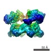 6205MC 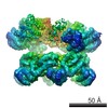 6204C 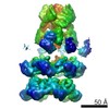 6206C 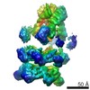 6207C 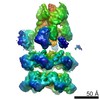 6208C 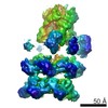 6209C 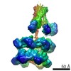 6210C 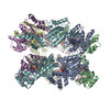 3j94C 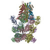 3j96C 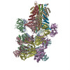 3j97C 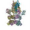 3j98C 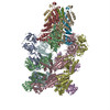 3j99C M: このデータのモデリングに利用したマップデータ C: 同じ文献を引用 ( |
|---|---|
| 類似構造データ |
-リンク
 PDBj
PDBj


- 集合体
集合体
| 登録構造単位 | 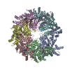
|
|---|---|
| 1 |
|
-要素
| #1: タンパク質 | / N-ethylmaleimide-sensitive fusion protein / NEM-sensitive fusion protein / Vesicular-fusion protein ...N-ethylmaleimide-sensitive fusion protein / NEM-sensitive fusion protein / Vesicular-fusion protein NSF / N-ethylmaleimide sensitive factor 分子量: 82907.430 Da / 分子数: 6 / 由来タイプ: 組換発現 由来: (組換発現) Cricetulus griseus (モンゴルキヌゲネズミ)遺伝子: NSF / 発現宿主:  Escherichia coli (大腸菌) / 参照: UniProt: P18708, vesicle-fusing ATPase Escherichia coli (大腸菌) / 参照: UniProt: P18708, vesicle-fusing ATPase#2: 化合物 | ChemComp-ADP / アデノシン二リン酸  分子量: 427.201 Da / 分子数: 4 / 由来タイプ: 合成 / 式: C10H15N5O10P2 / コメント: ADP, エネルギー貯蔵分子*YM 分子量: 427.201 Da / 分子数: 4 / 由来タイプ: 合成 / 式: C10H15N5O10P2 / コメント: ADP, エネルギー貯蔵分子*YM |
|---|
-実験情報
-実験
| 実験 | 手法: 電子顕微鏡法 |
|---|---|
| EM実験 | 試料の集合状態: PARTICLE / 3次元再構成法: 単粒子再構成法 |
- 試料調製
試料調製
| 構成要素 | 名称: ADP-bound N-ethylmaleimide sensitive factor / タイプ: COMPLEX / 詳細: hexamer |
|---|---|
| 分子量 | 値: 0.5 MDa / 実験値: NO |
| 緩衝液 | 名称: 50 mM Tris-Cl, 150 mM NaCl, 1 mM EDTA, 1 mM ATP, 1 mM DTT, 0.05% v/v Nonident P-40 pH: 8 詳細: 50 mM Tris-Cl, 150 mM NaCl, 1 mM EDTA, 1 mM ATP, 1 mM DTT, 0.05% v/v Nonident P-40 |
| 試料 | 濃度: 15 mg/ml / 包埋: NO / シャドウイング: NO / 染色: NO / 凍結: YES |
| 試料支持 | 詳細: Holey carbon on top of 400 mesh copper grid |
| 急速凍結 | 装置: FEI VITROBOT MARK I / 凍結剤: ETHANE / Temp: 90 K / 湿度: 100 % 詳細: Blot for 3.5 seconds before plunging into liquid ethane (FEI VITROBOT MARK I). 手法: Blot for 3.5 seconds before plunging |
- 電子顕微鏡撮影
| 実験機器 |  モデル: Tecnai Polara / 画像提供: FEI Company |
|---|---|
| 顕微鏡 | モデル: FEI POLARA 300 / 日付: 2014年1月14日 |
| 電子銃 | 電子線源: FIELD EMISSION GUN / 加速電圧: 300 kV / 照射モード: FLOOD BEAM |
| 電子レンズ | モード: BRIGHT FIELDBright-field microscopy / 倍率(公称値): 31000 X / 最大 デフォーカス(公称値): -2800 nm / 最小 デフォーカス(公称値): -1800 nm / Cs: 2.3 mm / カメラ長: 0 mm |
| 試料ホルダ | 試料ホルダーモデル: OTHER / 資料ホルダタイプ: unspecified |
| 撮影 | 電子線照射量: 44 e/Å2 / フィルム・検出器のモデル: GATAN K2 (4k x 4k) |
| 放射 | プロトコル: SINGLE WAVELENGTH / 単色(M)・ラウエ(L): M / 散乱光タイプ: x-ray |
| 放射波長 | 相対比: 1 |
- 解析
解析
| EMソフトウェア |
| |||||||||||||||||||||||||||||||||||||||||||||||||||||||||||||||||||||||||||||||||||||||||||||||||||||||||||||||||||||||||||||||||||||||||||||||||||||||||||||||||||||||||||||||||||||||||||||||||||||||||||||||||||||||||
|---|---|---|---|---|---|---|---|---|---|---|---|---|---|---|---|---|---|---|---|---|---|---|---|---|---|---|---|---|---|---|---|---|---|---|---|---|---|---|---|---|---|---|---|---|---|---|---|---|---|---|---|---|---|---|---|---|---|---|---|---|---|---|---|---|---|---|---|---|---|---|---|---|---|---|---|---|---|---|---|---|---|---|---|---|---|---|---|---|---|---|---|---|---|---|---|---|---|---|---|---|---|---|---|---|---|---|---|---|---|---|---|---|---|---|---|---|---|---|---|---|---|---|---|---|---|---|---|---|---|---|---|---|---|---|---|---|---|---|---|---|---|---|---|---|---|---|---|---|---|---|---|---|---|---|---|---|---|---|---|---|---|---|---|---|---|---|---|---|---|---|---|---|---|---|---|---|---|---|---|---|---|---|---|---|---|---|---|---|---|---|---|---|---|---|---|---|---|---|---|---|---|---|---|---|---|---|---|---|---|---|---|---|---|---|---|---|---|---|
| CTF補正 | 詳細: Each particle | |||||||||||||||||||||||||||||||||||||||||||||||||||||||||||||||||||||||||||||||||||||||||||||||||||||||||||||||||||||||||||||||||||||||||||||||||||||||||||||||||||||||||||||||||||||||||||||||||||||||||||||||||||||||||
| 対称性 | 点対称性: C1 (非対称) | |||||||||||||||||||||||||||||||||||||||||||||||||||||||||||||||||||||||||||||||||||||||||||||||||||||||||||||||||||||||||||||||||||||||||||||||||||||||||||||||||||||||||||||||||||||||||||||||||||||||||||||||||||||||||
| 3次元再構成 | 解像度: 7.6 Å / 解像度の算出法: FSC 0.143 CUT-OFF / 粒子像の数: 12830 / ピクセルサイズ(公称値): 2.4312 Å / ピクセルサイズ(実測値): 2.4312 Å / 詳細: (Single particle--Applied symmetry: C1) / 対称性のタイプ: POINT | |||||||||||||||||||||||||||||||||||||||||||||||||||||||||||||||||||||||||||||||||||||||||||||||||||||||||||||||||||||||||||||||||||||||||||||||||||||||||||||||||||||||||||||||||||||||||||||||||||||||||||||||||||||||||
| 原子モデル構築 | プロトコル: FLEXIBLE FIT / 空間: RECIPROCAL / Target criteria: R-factor 詳細: REFINEMENT PROTOCOL--flexible DETAILS--D2 domain of NSF was from crystal structure 1NSF. D1 domain of NSF was from related entry EMD-6204. | |||||||||||||||||||||||||||||||||||||||||||||||||||||||||||||||||||||||||||||||||||||||||||||||||||||||||||||||||||||||||||||||||||||||||||||||||||||||||||||||||||||||||||||||||||||||||||||||||||||||||||||||||||||||||
| 原子モデル構築 | PDB-ID: 1NSF PDB chain-ID: A / Accession code: 1NSF / Source name: PDB / タイプ: experimental model | |||||||||||||||||||||||||||||||||||||||||||||||||||||||||||||||||||||||||||||||||||||||||||||||||||||||||||||||||||||||||||||||||||||||||||||||||||||||||||||||||||||||||||||||||||||||||||||||||||||||||||||||||||||||||
| 精密化 | 解像度: 7.6→7.6 Å / SU ML: 1.3 / σ(F): 0.29 / 位相誤差: 36.78 / 立体化学のターゲット値: ML
| |||||||||||||||||||||||||||||||||||||||||||||||||||||||||||||||||||||||||||||||||||||||||||||||||||||||||||||||||||||||||||||||||||||||||||||||||||||||||||||||||||||||||||||||||||||||||||||||||||||||||||||||||||||||||
| 溶媒の処理 | 減衰半径: 0.9 Å / VDWプローブ半径: 1.11 Å / 溶媒モデル: FLAT BULK SOLVENT MODEL | |||||||||||||||||||||||||||||||||||||||||||||||||||||||||||||||||||||||||||||||||||||||||||||||||||||||||||||||||||||||||||||||||||||||||||||||||||||||||||||||||||||||||||||||||||||||||||||||||||||||||||||||||||||||||
| 原子変位パラメータ | Biso max: 50 Å2 / Biso mean: 50 Å2 / Biso min: 50 Å2 | |||||||||||||||||||||||||||||||||||||||||||||||||||||||||||||||||||||||||||||||||||||||||||||||||||||||||||||||||||||||||||||||||||||||||||||||||||||||||||||||||||||||||||||||||||||||||||||||||||||||||||||||||||||||||
| 精密化ステップ | サイクル: LAST / 解像度: 7.601→311.194 Å
| |||||||||||||||||||||||||||||||||||||||||||||||||||||||||||||||||||||||||||||||||||||||||||||||||||||||||||||||||||||||||||||||||||||||||||||||||||||||||||||||||||||||||||||||||||||||||||||||||||||||||||||||||||||||||
| 拘束条件 |
| |||||||||||||||||||||||||||||||||||||||||||||||||||||||||||||||||||||||||||||||||||||||||||||||||||||||||||||||||||||||||||||||||||||||||||||||||||||||||||||||||||||||||||||||||||||||||||||||||||||||||||||||||||||||||
| LS精密化 シェル | Refine-ID: ELECTRON MICROSCOPY / Total num. of bins used: 30
|