 ムービー
ムービー コントローラー


+ データを開く

- 基本情報
基本情報
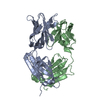
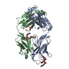
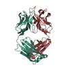
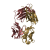
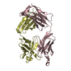
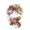
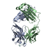
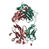
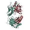
| 登録情報 | データベース: PDB / ID: 3gk8 | ||||||
|---|---|---|---|---|---|---|---|
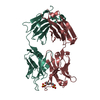
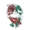
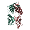
| タイトル | X-ray crystal structure of the Fab from MAb 14, mouse antibody against Canine Parvovirus | ||||||
 要素 要素 |
| ||||||
 キーワード キーワード |  IMMUNE SYSTEM (免疫系) / antibody fragment from neutralizing monoclonal antibody against canine parvovirus IMMUNE SYSTEM (免疫系) / antibody fragment from neutralizing monoclonal antibody against canine parvovirus | ||||||
| 機能・相同性 | 抗体 / Immunoglobulin-like / サンドイッチ / Mainly Beta 機能・相同性情報 機能・相同性情報 | ||||||
| 生物種 |  Mus musculus (ハツカネズミ) Mus musculus (ハツカネズミ) | ||||||
| 手法 | X線回折 / シンクロトロン / 分子置換 / 解像度: 2 Å | ||||||
 データ登録者 データ登録者 | Hafenstein, S. / Bowman, V. / Sun, T. / Nelson, C. / Palermo, L. / Chipman, P. / Battisti, A. / Parrish, C. | ||||||
 引用 引用 | ジャーナル: J Virol / 年: 2009 タイトル: Structural comparison of different antibodies interacting with parvovirus capsids. 著者: Susan Hafenstein / Valorie D Bowman / Tao Sun / Christian D S Nelson / Laura M Palermo / Paul R Chipman / Anthony J Battisti / Colin R Parrish / Michael G Rossmann /  要旨: The structures of canine parvovirus (CPV) and feline parvovirus (FPV) complexed with antibody fragments from eight different neutralizing monoclonal antibodies were determined by cryo-electron ...The structures of canine parvovirus (CPV) and feline parvovirus (FPV) complexed with antibody fragments from eight different neutralizing monoclonal antibodies were determined by cryo-electron microscopy (cryoEM) reconstruction to resolutions varying from 8.5 to 18 A. The crystal structure of one of the Fab molecules and the sequence of the variable domain for each of the Fab molecules have been determined. The structures of Fab fragments not determined crystallographically were predicted by homology modeling according to the amino acid sequence. Fitting of the Fab and virus structures into the cryoEM densities identified the footprints of each antibody on the viral surface. As anticipated from earlier analyses, the Fab binding sites are directed to two epitopes, A and B. The A site is on an exposed part of the surface near an icosahedral threefold axis, whereas the B site is about equidistant from the surrounding five-, three-, and twofold axes. One antibody directed to the A site binds CPV but not FPV. Two of the antibodies directed to the B site neutralize the virus as Fab fragments. The differences in antibody properties have been linked to the amino acids within the antibody footprints, the position of the binding site relative to the icosahedral symmetry elements, and the orientation of the Fab structure relative to the surface of the virus. Most of the exposed surface area was antigenic, although each of the antibodies had a common area of overlap that coincided with the positions of the previously mapped escape mutations. | ||||||
| 履歴 |
|
- 構造の表示
構造の表示
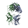
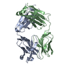
| 構造ビューア | 分子: MolmilJmol/JSmol |
|---|
- ダウンロードとリンク
-ダウンロード
| PDBx/mmCIF形式 | 3gk8.cif.gz | 92 KB | 表示 | PDBx/mmCIF形式 |
|---|---|---|---|---|
| PDB形式 | pdb3gk8.ent.gz | 73.6 KB | 表示 | PDB形式 |
| PDBx/mmJSON形式 | 3gk8.json.gz | ツリー表示 | PDBx/mmJSON形式 | |
| その他 |  その他のダウンロード その他のダウンロード |
-検証レポート
| アーカイブディレクトリ | https://data.pdbj.org/pub/pdb/validation_reports/gk/3gk8ftp://data.pdbj.org/pub/pdb/validation_reports/gk/3gk8 | HTTPS FTP |
|---|
-関連構造データ
| 関連構造データ | 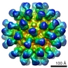 5105C 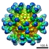 5106C 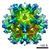 5107C 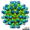 5108C 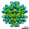 5109C 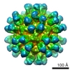 5110C 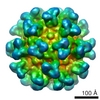 5111C 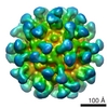 5112C  3iy0C  3iy1C  3iy2C  3iy3C 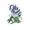 3iy4C  3iy5C  3iy6C  3iy7C C: 同じ文献を引用 ( |
|---|---|
| 類似構造データ |


-リンク
 PDBj
PDBj

- 集合体
集合体
| 登録構造単位 | 
| ||||||||
|---|---|---|---|---|---|---|---|---|---|
| 1 |
| ||||||||
| 単位格子 |
|
-要素
| #1: 抗体 | Fragment antigen-binding 分子量: 23261.770 Da / 分子数: 1 / 由来タイプ: 組換発現 / 由来: (組換発現) Mus musculus (ハツカネズミ) / Cell (発現宿主): Hybridoma |
|---|---|
| #2: 抗体 | Fragment antigen-binding 分子量: 23349.105 Da / 分子数: 1 / 由来タイプ: 組換発現 / 由来: (組換発現) Mus musculus (ハツカネズミ) / Cell (発現宿主): Hybridoma |
| #3: 水 | ChemComp-HOH / 水 分子量: 18.015 Da / 分子数: 113 / 由来タイプ: 天然 / 式: H2O 分子量: 18.015 Da / 分子数: 113 / 由来タイプ: 天然 / 式: H2O |
-実験情報
-実験
| 実験 | 手法: X線回折 / 使用した結晶の数: 1 |
|---|
- 試料調製
試料調製
| 結晶 | マシュー密度: 2.54 Å3/Da / 溶媒含有率: 51.64 % |
|---|---|
| 結晶化 | 温度: 298 K / 手法: 蒸気拡散法, ハンギングドロップ法 / pH: 7.5 詳細: 25% PEG 5000, 0.1M HEPES, pH 7.5, vapor diffusion, hanging drop, temperature 298K |
-データ収集
| 放射光源 | 由来: シンクロトロン / サイト: APS / ビームライン: 14-BM-D / 波長: 1 Å | |||||||||||||||||||||||||||||||||||||||||||||||||||||||||||||||||||||||||||||
|---|---|---|---|---|---|---|---|---|---|---|---|---|---|---|---|---|---|---|---|---|---|---|---|---|---|---|---|---|---|---|---|---|---|---|---|---|---|---|---|---|---|---|---|---|---|---|---|---|---|---|---|---|---|---|---|---|---|---|---|---|---|---|---|---|---|---|---|---|---|---|---|---|---|---|---|---|---|---|
| 検出器 | 日付: 2005年5月2日 | |||||||||||||||||||||||||||||||||||||||||||||||||||||||||||||||||||||||||||||
| 放射 | プロトコル: SINGLE WAVELENGTH / 散乱光タイプ: x-ray | |||||||||||||||||||||||||||||||||||||||||||||||||||||||||||||||||||||||||||||
| 放射波長 | 波長: 1 Å / 相対比: 1 | |||||||||||||||||||||||||||||||||||||||||||||||||||||||||||||||||||||||||||||
| Reflection | 冗長度: 3.6 % / Av σ(I) over netI: 25.82 / 数: 147415 / Rmerge(I) obs: 0.061 / Χ2: 1.39 / D res high: 1.85 Å / D res low: 50 Å / Num. obs: 40514 / % possible obs: 99.8 | |||||||||||||||||||||||||||||||||||||||||||||||||||||||||||||||||||||||||||||
| Diffraction reflection shell |
| |||||||||||||||||||||||||||||||||||||||||||||||||||||||||||||||||||||||||||||
| 反射 | 解像度: 1.85→50 Å / Num. obs: 40514 / % possible obs: 99.8 % / 冗長度: 3.6 % / Rmerge(I) obs: 0.061 / Χ2: 1.389 / Net I/σ(I): 25.819 | |||||||||||||||||||||||||||||||||||||||||||||||||||||||||||||||||||||||||||||
| 反射 シェル |
|
-位相決定
| 位相決定 | 手法: 分子置換 | ||||||
|---|---|---|---|---|---|---|---|
| Phasing MR | Rfactor: 0.447 / Cor.coef. Fo:Fc: 0.449 /
|
- 解析
解析
| ソフトウェア |
| ||||||||||||||||||||||||||||||||||||
|---|---|---|---|---|---|---|---|---|---|---|---|---|---|---|---|---|---|---|---|---|---|---|---|---|---|---|---|---|---|---|---|---|---|---|---|---|---|
| 精密化 | 構造決定の手法: 分子置換 / 解像度: 2→42.02 Å / Occupancy max: 1 / Occupancy min: 1 / FOM work R set: 0.756 /
| ||||||||||||||||||||||||||||||||||||
| 原子変位パラメータ | Biso max: 99.13 Å2 / Biso mean: 41.161 Å2 / Biso min: 1.89 Å2
| ||||||||||||||||||||||||||||||||||||
| 精密化ステップ | サイクル: LAST / 解像度: 2→42.02 Å
| ||||||||||||||||||||||||||||||||||||
| 拘束条件 |
|
