 ムービー
ムービー コントローラー


+ データを開く

- 基本情報
基本情報
| 登録情報 | データベース: PDB / ID: 3equ | ||||||
|---|---|---|---|---|---|---|---|
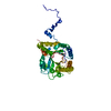
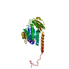
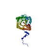
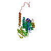
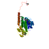
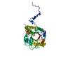
| タイトル | Crystal structure of penicillin-binding protein 2 from Neisseria gonorrhoeae | ||||||
 要素 要素 | Penicillin-binding protein 2 | ||||||
 キーワード キーワード |  BIOSYNTHETIC PROTEIN (生合成) / Penicillin-binding protein (ペニシリン結合タンパク質) / Class B transpeptidase / Cell division (細胞分裂) / Cell inner membrane / Cell membrane (細胞膜) / Cell shape / Cell wall biogenesis/degradation / Peptidoglycan synthesis (ペプチドグリカン) BIOSYNTHETIC PROTEIN (生合成) / Penicillin-binding protein (ペニシリン結合タンパク質) / Class B transpeptidase / Cell division (細胞分裂) / Cell inner membrane / Cell membrane (細胞膜) / Cell shape / Cell wall biogenesis/degradation / Peptidoglycan synthesis (ペプチドグリカン) | ||||||
| 機能・相同性 |  機能・相同性情報peptidoglycan glycosyltransferase activity / serine-type D-Ala-D-Ala carboxypeptidase / FtsZ-dependent cytokinesis / division septum assembly / serine-type D-Ala-D-Ala carboxypeptidase activity / penicillin binding / peptidoglycan biosynthetic process / cell wall organization / regulation of cell shape / response to antibiotic ...peptidoglycan glycosyltransferase activity / serine-type D-Ala-D-Ala carboxypeptidase / FtsZ-dependent cytokinesis / division septum assembly / serine-type D-Ala-D-Ala carboxypeptidase activity / penicillin binding / peptidoglycan biosynthetic process / cell wall organization / regulation of cell shape / response to antibiotic / タンパク質分解 / 細胞膜 機能・相同性情報peptidoglycan glycosyltransferase activity / serine-type D-Ala-D-Ala carboxypeptidase / FtsZ-dependent cytokinesis / division septum assembly / serine-type D-Ala-D-Ala carboxypeptidase activity / penicillin binding / peptidoglycan biosynthetic process / cell wall organization / regulation of cell shape / response to antibiotic ...peptidoglycan glycosyltransferase activity / serine-type D-Ala-D-Ala carboxypeptidase / FtsZ-dependent cytokinesis / division septum assembly / serine-type D-Ala-D-Ala carboxypeptidase activity / penicillin binding / peptidoglycan biosynthetic process / cell wall organization / regulation of cell shape / response to antibiotic / タンパク質分解 / 細胞膜類似検索 - 分子機能 | ||||||
| 生物種 |  Neisseria gonorrhoeae (淋菌) Neisseria gonorrhoeae (淋菌) | ||||||
| 手法 | X線回折 / シンクロトロン / 解像度: 2.4 Å | ||||||
 データ登録者 データ登録者 | Powell, A.J. / Deacon, A.M. / Nicholas, R.A. / Davies, C. | ||||||
 引用 引用 | ジャーナル: J.Biol.Chem. / 年: 2009 タイトル: Crystal Structures of Penicillin-binding Protein 2 from Penicillin-susceptible and -resistant Strains of Neisseria gonorrhoeae Reveal an Unexpectedly Subtle Mechanism for Antibiotic Resistance. 著者: Powell, A.J. / Tomberg, J. / Deacon, A.M. / Nicholas, R.A. / Davies, C. | ||||||
| 履歴 |
|
- 構造の表示
構造の表示
| 構造ビューア | 分子: MolmilJmol/JSmol |
|---|
- ダウンロードとリンク
-ダウンロード
| PDBx/mmCIF形式 | 3equ.cif.gz | 179.9 KB | 表示 | PDBx/mmCIF形式 |
|---|---|---|---|---|
| PDB形式 | pdb3equ.ent.gz | 141 KB | 表示 | PDB形式 |
| PDBx/mmJSON形式 | 3equ.json.gz | ツリー表示 | PDBx/mmJSON形式 | |
| その他 |  その他のダウンロード その他のダウンロード |
-検証レポート
| アーカイブディレクトリ | https://data.pdbj.org/pub/pdb/validation_reports/eq/3equftp://data.pdbj.org/pub/pdb/validation_reports/eq/3equ | HTTPS FTP |
|---|
-関連構造データ


-リンク
 PDBj
PDBj
- 集合体
集合体
| 登録構造単位 | 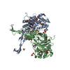
| ||||||||
|---|---|---|---|---|---|---|---|---|---|
| 1 | 
| ||||||||
| 2 | 
| ||||||||
| 単位格子 |
|
-要素
| #1: タンパク質 | 分子量: 59607.934 Da / 分子数: 2 / 由来タイプ: 組換発現 / 由来: (組換発現) Neisseria gonorrhoeae (淋菌) / 株: FA19 / 遺伝子: penA / プラスミド: pMAL-C2KV/H6 / 発現宿主: Escherichia coli (大腸菌) / 株 (発現宿主): GW6011参照: UniProt: P08149, serine-type D-Ala-D-Ala carboxypeptidase#2: 化合物 | ChemComp-SO4 / 硫酸塩  分子量: 96.063 Da / 分子数: 17 / 由来タイプ: 合成 / 式: SO4 分子量: 96.063 Da / 分子数: 17 / 由来タイプ: 合成 / 式: SO4#3: 化合物 | グリセリン  分子量: 92.094 Da / 分子数: 2 / 由来タイプ: 合成 / 式: C3H8O3 分子量: 92.094 Da / 分子数: 2 / 由来タイプ: 合成 / 式: C3H8O3#4: 水 | ChemComp-HOH / | 水 分子量: 18.015 Da / 分子数: 146 / 由来タイプ: 天然 / 式: H2O 分子量: 18.015 Da / 分子数: 146 / 由来タイプ: 天然 / 式: H2O |
|---|
-実験情報
-実験
| 実験 | 手法: X線回折 / 使用した結晶の数: 1 |
|---|
- 試料調製
試料調製
| 結晶 | マシュー密度: 3.8 Å3/Da / 溶媒含有率: 67.66 % |
|---|---|
| 結晶化 | 温度: 294 K / 手法: 蒸気拡散法, ハンギングドロップ法 / pH: 8.5 詳細: 2.2 M ammonium sulphate, 100 mM Tris-HCl, pH 8.5, VAPOR DIFFUSION, HANGING DROP, temperature 294K |
-データ収集
| 回折 | 平均測定温度: 100 K |
|---|---|
| 放射光源 | 由来: シンクロトロン / サイト: SSRL  / ビームライン: BL9-2 / 波長: 1 Å / ビームライン: BL9-2 / 波長: 1 Å |
| 検出器 | タイプ: ADSC QUANTUM 4 / 検出器: CCD / 日付: 2001年3月20日 |
| 放射 | モノクロメーター: Si (111) / プロトコル: SINGLE WAVELENGTH / 単色(M)・ラウエ(L): M / 散乱光タイプ: x-ray |
| 放射波長 | 波長: 1 Å / 相対比: 1 |
| 反射 | 解像度: 2.3→87 Å / Num. all: 73860 / Num. obs: 73860 / % possible obs: 90.3 % / Observed criterion σ(F): 0 / Observed criterion σ(I): 0 / 冗長度: 6.2 % / Biso Wilson estimate: 43.2 Å2 / Rsym value: 0.061 / Net I/σ(I): 8.8 |
| 反射 シェル | 解像度: 2.3→2.36 Å / 冗長度: 3.7 % / Mean I/σ(I) obs: 0 / Num. unique all: 3081 / Rsym value: 0.349 / % possible all: 52.6 |
- 解析
解析
| ソフトウェア |
| ||||||||||||||||||||||||||||||||||||||||||||||||||||||||||||||||||||||||||||||||||||||||||
|---|---|---|---|---|---|---|---|---|---|---|---|---|---|---|---|---|---|---|---|---|---|---|---|---|---|---|---|---|---|---|---|---|---|---|---|---|---|---|---|---|---|---|---|---|---|---|---|---|---|---|---|---|---|---|---|---|---|---|---|---|---|---|---|---|---|---|---|---|---|---|---|---|---|---|---|---|---|---|---|---|---|---|---|---|---|---|---|---|---|---|---|
| 精密化 | 解像度: 2.4→66.6 Å / Cor.coef. Fo:Fc: 0.93 / Cor.coef. Fo:Fc free: 0.914 / SU B: 10.749 / SU ML: 0.126 / Isotropic thermal model: isotropic / 交差検証法: THROUGHOUT / σ(F): 0 / ESU R: 0.215 / ESU R Free: 0.187 / 立体化学のターゲット値: MAXIMUM LIKELIHOOD
| ||||||||||||||||||||||||||||||||||||||||||||||||||||||||||||||||||||||||||||||||||||||||||
| 溶媒の処理 | イオンプローブ半径: 0.8 Å / 減衰半径: 0.8 Å / VDWプローブ半径: 1.2 Å / 溶媒モデル: MASK | ||||||||||||||||||||||||||||||||||||||||||||||||||||||||||||||||||||||||||||||||||||||||||
| 原子変位パラメータ | Biso mean: 36.7 Å2
| ||||||||||||||||||||||||||||||||||||||||||||||||||||||||||||||||||||||||||||||||||||||||||
| 精密化ステップ | サイクル: LAST / 解像度: 2.4→66.6 Å
| ||||||||||||||||||||||||||||||||||||||||||||||||||||||||||||||||||||||||||||||||||||||||||
| 拘束条件 |
| ||||||||||||||||||||||||||||||||||||||||||||||||||||||||||||||||||||||||||||||||||||||||||
| LS精密化 シェル | 解像度: 2.4→2.462 Å / Total num. of bins used: 20
|
