 ムービー
ムービー コントローラー


+ データを開く

- 基本情報
基本情報
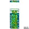
| 登録情報 | データベース: PDB / ID: 3dtp | ||||||
|---|---|---|---|---|---|---|---|
| タイトル | Tarantula heavy meromyosin obtained by flexible docking to Tarantula muscle thick filament Cryo-EM 3D-MAP | ||||||
 要素 要素 |
| ||||||
 キーワード キーワード |  CONTRACTILE PROTEIN / MUSCLE PROTEIN (骨格筋) / SMOOTH MUSCLE (平滑筋) / MYOSIN SUBFRAGMENT 2 / HEAVY MEROMYOSIN / ESSENTIAL LIGHT CHAIN / REGULATORY LIGHT CHAIN / MOTOR PROTEIN (モータータンパク質) / COILED-COIL (コイルドコイル) CONTRACTILE PROTEIN / MUSCLE PROTEIN (骨格筋) / SMOOTH MUSCLE (平滑筋) / MYOSIN SUBFRAGMENT 2 / HEAVY MEROMYOSIN / ESSENTIAL LIGHT CHAIN / REGULATORY LIGHT CHAIN / MOTOR PROTEIN (モータータンパク質) / COILED-COIL (コイルドコイル) | ||||||
| 機能・相同性 |  機能・相同性情報 機能・相同性情報myosin II filament / regulation of slow-twitch skeletal muscle fiber contraction / regulation of the force of skeletal muscle contraction / myofibril assembly / elastic fiber assembly / myosin light chain binding / skeletal muscle myosin thick filament assembly / muscle myosin complex / myosin II binding / muscle filament sliding ...myosin II filament / regulation of slow-twitch skeletal muscle fiber contraction / regulation of the force of skeletal muscle contraction / myofibril assembly / elastic fiber assembly / myosin light chain binding / skeletal muscle myosin thick filament assembly / muscle myosin complex / myosin II binding / muscle filament sliding / regulation of the force of heart contraction / ミオフィラメント / transition between fast and slow fiber / myosin filament / actomyosin structure organization / myosin II complex / adult heart development / cardiac muscle hypertrophy in response to stress / cardiac muscle cell development / myosin complex / structural constituent of muscle / sarcomere organization / microfilament motor activity / ventricular cardiac muscle tissue morphogenesis / myofibril / myosin heavy chain binding / skeletal muscle contraction / smooth muscle contraction / striated muscle contraction / stress fiber / ATP metabolic process / cardiac muscle contraction / regulation of heart rate / sarcomere / muscle contraction / ADP binding / Z disc / actin filament binding / actin binding / calmodulin binding / calcium ion binding / magnesium ion binding / ATP binding / 細胞質基質 / 細胞質類似検索 - 分子機能 | ||||||
| 生物種 |  Gallus gallus (ニワトリ) Gallus gallus (ニワトリ) Homo sapiens (ヒト) Homo sapiens (ヒト) Avicularia avicularia (クモ) Avicularia avicularia (クモ) | ||||||
| 手法 | 電子顕微鏡法 / らせん対称体再構成法 / クライオ電子顕微鏡法 / 解像度: 20 Å | ||||||
 データ登録者 データ登録者 | Alamo, L. / Wriggers, W. / Pinto, A. / Bartoli, F. / Salazar, L. / Zhao, F.Q. / Craig, R. / Padron, R. | ||||||
 引用 引用 | ジャーナル: J Mol Biol / 年: 2008 タイトル: Three-dimensional reconstruction of tarantula myosin filaments suggests how phosphorylation may regulate myosin activity. 著者: Lorenzo Alamo / Willy Wriggers / Antonio Pinto / Fulvia Bártoli / Leiria Salazar / Fa-Qing Zhao / Roger Craig / Raúl Padrón /  要旨: Muscle contraction involves the interaction of the myosin heads of the thick filaments with actin subunits of the thin filaments. Relaxation occurs when this interaction is blocked by molecular ...Muscle contraction involves the interaction of the myosin heads of the thick filaments with actin subunits of the thin filaments. Relaxation occurs when this interaction is blocked by molecular switches on these filaments. In many muscles, myosin-linked regulation involves phosphorylation of the myosin regulatory light chains (RLCs). Electron microscopy of vertebrate smooth muscle myosin molecules (regulated by phosphorylation) has provided insight into the relaxed structure, revealing that myosin is switched off by intramolecular interactions between its two heads, the free head and the blocked head. Three-dimensional reconstruction of frozen-hydrated specimens revealed that this asymmetric head interaction is also present in native thick filaments of tarantula striated muscle. Our goal in this study was to elucidate the structural features of the tarantula filament involved in phosphorylation-based regulation. A new reconstruction revealed intra- and intermolecular myosin interactions in addition to those seen previously. To help interpret the interactions, we sequenced the tarantula RLC and fitted an atomic model of the myosin head that included the predicted RLC atomic structure and an S2 (subfragment 2) crystal structure to the reconstruction. The fitting suggests one intramolecular interaction, between the cardiomyopathy loop of the free head and its own S2, and two intermolecular interactions, between the cardiac loop of the free head and the essential light chain of the blocked head and between the Leu305-Gln327 interaction loop of the free head and the N-terminal fragment of the RLC of the blocked head. These interactions, added to those previously described, would help switch off the thick filament. Molecular dynamics simulations suggest how phosphorylation could increase the helical content of the RLC N-terminus, weakening these interactions, thus releasing both heads and activating the thick filament. #1: ジャーナル: J Mol Biol / 年: 2003 タイトル: Refined model of the 10S conformation of smooth muscle myosin by cryo-electron microscopy 3D image reconstruction. 著者: Jun Liu / Thomas Wendt / Dianne Taylor / Kenneth Taylor /  要旨: The actin-activated ATPase activity of smooth muscle myosin and heavy meromyosin (smHMM) is regulated by phosphorylation of the regulatory light chain (RLC). Complete regulation requires two intact ...The actin-activated ATPase activity of smooth muscle myosin and heavy meromyosin (smHMM) is regulated by phosphorylation of the regulatory light chain (RLC). Complete regulation requires two intact myosin heads because single-headed myosin subfragments are always active. 2D crystalline arrays of the 10S form of intact myosin, which has a dephosphorylated RLC, were produced on a positively charged lipid monolayer and imaged in 3D at 2.0 nm resolution by cryo-electron microscopy of frozen, hydrated specimens. An atomic model of smooth muscle myosin was constructed from the X-ray structures of the smooth muscle myosin motor domain and essential light chain and a homology model of the RLC was produced based on the skeletal muscle S1 structure. The initial model of the 10S myosin, based on the previous reconstruction of smHMM, was subjected to real space refinement to obtain a quantitative fit to the density. The smHMM was likewise refined and both refined models reveal the same asymmetric interaction between the upper 50 kDa domain of the "blocked" head and parts of the catalytic, converter domains and the essential light chain of the "free" head observed previously. This observation suggests that this interaction is not simply due to crystallographic packing but is enforced by elements of the myosin heads. The 10S reconstruction shows additional alpha-helical coiled-coil not seen in the earlier smHMM reconstruction, but the location of one segment of S2 is the same in both. #2: ジャーナル: Proc Natl Acad Sci U S A / 年: 2006タイトル: Crystal structures of human cardiac beta-myosin II S2-Delta provide insight into the functional role of the S2 subfragment. 著者: Wulf Blankenfeldt / Nicolas H Thomä / John S Wray / Mathias Gautel / Ilme Schlichting /  要旨: Myosin II is the major component of the muscle thick filament. It consists of two N-terminal S1 subfragments ("heads") connected to a long dimeric coiled-coil rod. The rod is in itself twofold ...Myosin II is the major component of the muscle thick filament. It consists of two N-terminal S1 subfragments ("heads") connected to a long dimeric coiled-coil rod. The rod is in itself twofold symmetric, but in the filament, the two heads point away from the filament surface and are therefore not equivalent. This breaking of symmetry requires the initial section of the rod, subfragment 2 (S2), to be relatively flexible. S2 is an important functional element, involved in various mechanisms by which the activity of smooth and striated muscle is regulated. We have determined crystal structures of the 126 N-terminal residues of S2 from human cardiac beta-myosin II (S2-Delta), of both WT and the disease-associated E924K mutant. S2-Delta is a straight parallel dimeric coiled coil, but the N terminus of one chain is disordered in WT-S2-Delta due to crystal contacts, indicative of unstable local structure. Bulky noncanonical side chains pack into a/d positions of S2-Delta's N terminus, leading to defined local asymmetry and axial stagger, which could induce nonequivalence of the S1 subfragments. Additionally, S2 possesses a conserved charge distribution with three prominent rings of negative potential within S2-Delta, the first of which may provide a binding interface for the "blocked head" of smooth muscle myosin in the OFF state. The observation that many disease-associated mutations affect the second negatively charged ring further suggests that charge interactions play an important role in regulation of cardiac muscle activity through myosin-binding protein C. #3: ジャーナル: Cell / 年: 1999タイトル: Atomic structure of scallop myosin subfragment S1 complexed with MgADP: a novel conformation of the myosin head. 著者: A Houdusse / V N Kalabokis / D Himmel / A G Szent-Györgyi / C Cohen / 要旨: The crystal structure of a proteolytic subfragment from scallop striated muscle myosin, complexed with MgADP, has been solved at 2.5 A resolution and reveals an unusual conformation of the myosin ...The crystal structure of a proteolytic subfragment from scallop striated muscle myosin, complexed with MgADP, has been solved at 2.5 A resolution and reveals an unusual conformation of the myosin head. The converter and the lever arm are in very different positions from those in either the pre-power stroke or near-rigor state structures; moreover, in contrast to these structures, the SH1 helix is seen to be unwound. Here we compare the overall organization of the myosin head in these three states and show how the conformation of three flexible "joints" produces rearrangements of the four major subdomains in the myosin head with different bound nucleotides. We believe that this novel structure represents one of the prehydrolysis ("ATP") states of the contractile cycle in which the myosin heads stay detached from actin. | ||||||
| 履歴 |
|
- 構造の表示
構造の表示
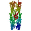
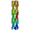
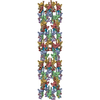
| ムービー |
ムービービューア |
|---|---|
| 構造ビューア | 分子: MolmilJmol/JSmol |
- ダウンロードとリンク
-ダウンロード
| PDBx/mmCIF形式 | 3dtp.cif.gz | 543.4 KB | 表示 | PDBx/mmCIF形式 |
|---|---|---|---|---|
| PDB形式 | pdb3dtp.ent.gz | 444.5 KB | 表示 | PDB形式 |
| PDBx/mmJSON形式 | 3dtp.json.gz | ツリー表示 | PDBx/mmJSON形式 | |
| その他 |  その他のダウンロード その他のダウンロード |
-検証レポート
| アーカイブディレクトリ | https://data.pdbj.org/pub/pdb/validation_reports/dt/3dtpftp://data.pdbj.org/pub/pdb/validation_reports/dt/3dtp | HTTPS FTP |
|---|
-関連構造データ
| 関連構造データ | 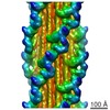 1950MU M: このデータのモデリングに利用したマップデータ U: あてはまらない、座標系が異なる*YM |
|---|---|
| 類似構造データ |
-リンク
 PDBj
PDBj


- 集合体
集合体
| 登録構造単位 | 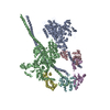
|
|---|---|
| 1 | x 36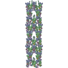
|
| 2 |
|
| 3 | 
|
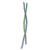
| 対称性 | らせん対称: (回転対称性: 4 / Dyad axis: no / N subunits divisor: 1 / Num. of operations: 36 / Rise per n subunits: 145 Å / Rotation per n subunits: 30 °) |
-要素
| #1: タンパク質 | 分子量: 111851.070 Da / 分子数: 1 断片: SUBFRAGMENT 1(S1), DELTA-S2 (residues 2-972),SUBFRAGMENT 1(S1), DELTA-S2 (residues 2-972) 由来タイプ: 組換発現 由来: (組換発現) Gallus gallus (ニワトリ), (組換発現) Homo sapiens (ヒト)遺伝子: MYH11, MYH7, MYHCB / プラスミド: PVL1392 / 細胞株 (発現宿主): SF9 発現宿主:  Spodoptera frugiperda (ツマジロクサヨトウ) Spodoptera frugiperda (ツマジロクサヨトウ)参照: UniProt: P10587, UniProt: P12883 | ||||
|---|---|---|---|---|---|
| #2: タンパク質 | 分子量: 112051.328 Da / 分子数: 1 断片: SUBFRAGMENT 1(S1), DELTA-S2 (residues 2-974),SUBFRAGMENT 1(S1), DELTA-S2 (residues 2-974) 由来タイプ: 組換発現 由来: (組換発現) Gallus gallus (ニワトリ), (組換発現) Homo sapiens (ヒト)遺伝子: MYH11, MYH7, MYHCB / プラスミド: PVL1392 / 細胞株 (発現宿主): SF9 発現宿主: Spodoptera frugiperda (ツマジロクサヨトウ)参照: UniProt: P10587, UniProt: P12883 | ||||
| #3: タンパク質 | 分子量: 16873.025 Da / 分子数: 2 / 由来タイプ: 組換発現 / 由来: (組換発現) Gallus gallus (ニワトリ) / 遺伝子: MYL6 / プラスミド: PVL1392 / 細胞株 (発現宿主): SF9発現宿主: Spodoptera frugiperda (ツマジロクサヨトウ)参照: UniProt: P02607 #4: タンパク質 | 分子量: 21709.250 Da / 分子数: 2 / 由来タイプ: 天然 / 由来: (天然) Avicularia avicularia (クモ) / 組織: leg muscle / 参照: UniProt: B4XT43配列の詳細 | THE CONFLICT LISTED FOR CHAINS A AND B, RESIDUE 2 ARE CONSISTENT WITH PDB ENTRY 1BR1 AND 1I84. THE ...THE CONFLICT LISTED FOR CHAINS A AND B, RESIDUE 2 ARE CONSISTENT | |
-実験情報
-実験
| 実験 | 手法: 電子顕微鏡法 |
|---|---|
| EM実験 | 試料の集合状態: FILAMENT / 3次元再構成法: らせん対称体再構成法 |
- 試料調製
試料調製
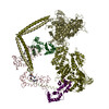
| 構成要素 | 名称: Myosin thick filaments from Tarantula striated muscle タイプ: COMPLEX 詳細: Polymer of a multiple myosin assembled over a paramyosin core. MODEL BUILDING: the atomic model consists of two s1 heads and a segment of s2. each heavy meromyosin consists of a chimera built ...詳細: Polymer of a multiple myosin assembled over a paramyosin core. MODEL BUILDING: the atomic model consists of two s1 heads and a segment of s2. each heavy meromyosin consists of a chimera built by chicken smooth muscle heavy chain (1i84) for s1 plus human cardiac muscle (2fxm) for s2 (chains a,b) and two light chains, the chicken smooth muscle (1i84) for elc (chains c,d) and a homology model based on 1br1 of the tarantula skeletal muscle rlc sequence (chains e,f), this model was flexible fitted to a tarantula 3d map (emd-1535) |
|---|---|
| 緩衝液 | 名称: 100mM NaCl, 3mM MgCl2, 1mM EGTA, 5mM PIPES, 5mM NaH2PO4, 1mM NaN3 pH: 7 詳細: 100mM NaCl, 3mM MgCl2, 1mM EGTA, 5mM PIPES, 5mM NaH2PO4, 1mM NaN3 |
| 試料 | 包埋: NO / シャドウイング: NO / 染色: NO / 凍結: YES |
| 試料支持 | 詳細: Holey carbon grids, 400 mesh |
| 急速凍結 | 詳細: Plunging in a liquid ethane. Blotting was performed from one side of the grid till a thin sample film on it using Whatman No 42 filter paper, then the grid was immediately plunged under ...詳細: Plunging in a liquid ethane. Blotting was performed from one side of the grid till a thin sample film on it using Whatman No 42 filter paper, then the grid was immediately plunged under gravity into liquid ethane cooled by liquid nitrogen. Grids were stored under liquid nitrogen. |
- 電子顕微鏡撮影
| 顕微鏡 | モデル: FEI/PHILIPS CM120T / 日付: 2001年9月19日 詳細: Holey carbon grids Cryo preserved in Liquid ethane were observed in a Philips CM120 electron microscope under low dose conditions. Only filaments on thin carbon over holes were photographed |
|---|---|
| 電子銃 | 電子線源: LAB6 / 加速電圧: 120 kV / 照射モード: FLOOD BEAM |
| 電子レンズ | モード: BRIGHT FIELDBright-field microscopy / 倍率(公称値): 35000 X / 倍率(補正後): 35000 X / 最大 デフォーカス(公称値): 1950 nm / 最小 デフォーカス(公称値): 1950 nm / Cs: 2 mm |
| 試料ホルダ | 温度: 88 K |
| 画像スキャン | デジタル画像の数: 465 |
- 解析
解析
| EMソフトウェア |
| ||||||||||||
|---|---|---|---|---|---|---|---|---|---|---|---|---|---|
| CTF補正 | 詳細: no corrected | ||||||||||||
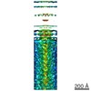
| 3次元再構成 | 手法: Single particle reconstruction with a modification of the IHRSR method 解像度: 20 Å / 粒子像の数: 15504 / ピクセルサイズ(公称値): 2.48 Å / ピクセルサイズ(実測値): 2.48 Å / 倍率補正: Tropomyosin paracrystal 詳細: three-dimensional single particle reconstruction was carried out by a modification of the ihrsr method, using spider. low-dose electron micrographs of 1008 frozen-hydrated thick filaments ...詳細: three-dimensional single particle reconstruction was carried out by a modification of the ihrsr method, using spider. low-dose electron micrographs of 1008 frozen-hydrated thick filaments halves were digitized at 0.248 nm per pixel using a nikon super coolscan 8000 ed scanner. filaments were aligned with the bare zone at the top, to ensure correct polarity in subsequent steps. a total of 15,504 segments, each 62 nm long, with an overlap of 55.8 nm, and containing aprox. 40,000 unique pairs of interacting myosin heads went into the reconstruction. as an initial reference model we used the tarantula negatively stained 3d-map, which was axially rotated, axially shifted and also out of plane tilted up to plus-minus12deg. for projection matching, giving a total of 4,095 projections (13 tilted projections plus-minus 12 deg. every 2 deg., 45 reference rotated projections (0-90 deg., every 2 deg. rotation angle), and 7 image axial shifts of 2.2 nm. the resulting 3d-map combines about 10,700 out of 15,504 filament segments, a yield of 69 percent of included segments. 対称性のタイプ: HELICAL | ||||||||||||
| 原子モデル構築 | プロトコル: FLEXIBLE FIT / 空間: REAL / Target criteria: Constrained Molecular Dynamics 詳細: METHOD--Flexible Fitting REFINEMENT PROTOCOL--Custom skeleton of 31 positional markers | ||||||||||||
| 原子モデル構築 |
| ||||||||||||
| 精密化ステップ | サイクル: LAST
|
